Review
. 2021 Jan;10(1):28-42.
doi: 10.21037/acs-2020-mv-16.

Imaging the mitral valve: a primer for the interventional surgeon

Affiliations
Review

Imaging the mitral valve: a primer for the interventional surgeon

Aditya Sengupta et al. Ann Cardiothorac Surg. 2021 Jan.

Abstract

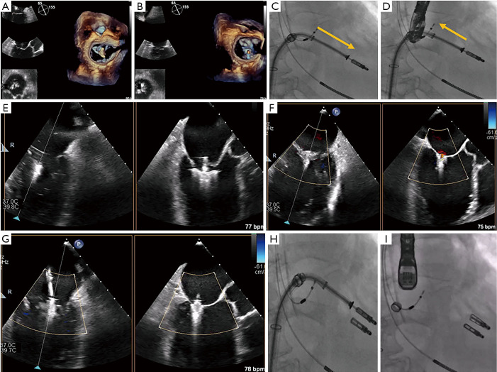
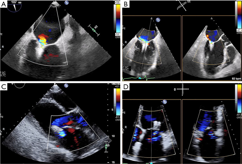
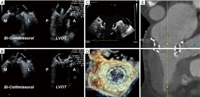
Transcatheter mitral valve interventions (TMVI) have evolved over the past decade as alternatives to open surgical repair for the therapeutic management of patients with severe mitral regurgitation (MR). Concurrent with the development of these technologies, quality multi-modality cardiac imaging has become essential in patient selection and procedural guidance. The former involves assessments of the pathophysiologic mechanisms of regurgitation, valvular anatomy and morphology, as well as objective quantification of the severity of MR. Both transthoracic and transesophageal echocardiography (TEE) are crucial and serve as the gateway to diagnosis and management of mitral valvular disease. Along with multi-detector computed tomography (CT) and cardiac magnetic resonance imaging (CMR), echocardiography plays an important role for preprocedural planning and evaluation of the spatial relationships of the mitral valvular complex with the coronary sinus, circumflex coronary artery and left ventricular (LV) outflow tract. Procedures that target mitral leaflets (e.g., MitraClip, PASCAL) or annulus (e.g., Cardioband, Carillon), or provide chordal (e.g., NeoChord, Harpoon) or valvular replacement, tend to be guided by TEE and assisted by fluoroscopy. As newer devices become available and outcomes of TMVI improve, cardiac imaging will undoubtedly continue to play an essential role in the success of percutaneous mitral valve repair (MVr) and replacement. The interventional surgeon of the future must therefore have a thorough understanding of the various imaging modalities while synthesizing and integrating novel concepts (e.g., neo-LV outflow tract) as applicable to assessing valvular function and pathology.

Keywords: Mitral regurgitation (MR); computed tomography (CT); interventional echocardiography; multimodality cardiac imaging; transcatheter mitral valve replacement (TMVR).


Conflict of interest statement

Conflicts of Interest: Dr. GHLT is a consultant for Medtronic, Abbott Structural Heart, and W. L. Gore & Associates. Dr. RPM reports the following financial interests/arrangements or affiliations: Steering Committee, Abbott TRILUMINATE Pivotal; Speakers Bureau, Edwards Lifesciences; Speakers Bureau, Medtronic Heart Valves; Medical Advisory Board, CardioCare-Edwards; Executive Committee, Medtronic TMVR, Officer and Stock Holder, Bay Labs-AI Company. The other authors have no conflicts of interest to declare.

Figures

Figure 1
Figure 1
Pathophysiology of mitral regurgitation. Ascertaining the pathophysiologic mechanism(s) of MR is a key step in evaluating patients for TMVI. (A,B) Primary MR occurs as a result of degenerative disease affecting one or more components of the mitral valvular complex. A large A2-A3 flail anterior leaflet (star) with torrential MR is demonstrated. (C,D) In contrast, secondary MR is a consequence of left ventricular dilatation and remodeling, which leads to leaflet tethering and failure of coaptation. Echocardiography here shows severe functional MR in a patient with an ejection fraction of 15%. A2, middle segment of the anterior mitral leaflet; A3, posterior segment of the anterior mitral leaflet; MR, mitral regurgitation; TMVI, transcatheter mitral valve intervention.
Figure 2
Figure 2
Morphological analysis of the mitral valve in primary mitral regurgitation. Preprocedural TEE is often useful in distinguishing between Barlow’s disease and fibroelastic deficiency in patients with primary MR. (A,B) The four-chamber view shows a flail A1 segment (star) of the anterior leaflet and severe MR. (C,D) A flail P2 scallop (star) with severe MR is seen on the AV long-axis view. (E,F) TEE here shows torrential MR due to lateral commissural prolapse extending to the P1 and P2 segments (star), with billowing well into the left atrium. A1, anterior segment of the anterior mitral leaflet; AV, aortic valve; MR, mitral regurgitation; P1, anterior segment of the posterior mitral leaflet; P2, middle segment of the posterior mitral leaflet; TEE, transesophageal echocardiography.
Figure 3
Figure 3
Preprocedural anatomic assessment with transthoracic echocardiography. TTE remains the cornerstone of anatomic evaluation in patients with mitral valve disease. (A) The apical four-chamber view visualizes both mitral leaflets and their coaptation, which should be at the level of the mitral annulus. Here, we see PML prolapse. (B) Color Doppler imaging reveals moderate to severe MR with an anteriorly directed jet. (C) The parasternal long-axis view allows for assessment of leaflet thickness and motion during the cardiac cycle. The left ventricular end-systolic dimension here is measured to be 2.8 cm. (D) The apical two-chamber view is essential for evaluating the different segments of the mitral leaflets and the severity of MR. Here, the LA volume index was measured at 60 mL/m2. AML, anterior mitral leaflet; LA, left atrium; MR, mitral regurgitation; PML, posterior mitral leaflet; TTE, transthoracic echocardiography.
Figure 4
Figure 4
Preprocedural multidetector computed tomography analysis. Using the 3Mensio Valves software, MDCT reconstruction can be used for preprocedural evaluation of valvular anatomy to determine the feasibility of TMVR. (A) First, annular dimensions (intercommissural maximal diameter and anterior-posterior diameter) are determined. (B) Next, the neo-LVOT area is measured by overlaying a virtual TMVR device in the appropriate position (1.86 cm2 in this case). (C) The virtually-placed TMVR device across the mitral annulus is used to assess LVOT clearance in a 3-chamber view. (D) The white line cutting across the LVOT and virtual TMVR device demarcates the cut-plane used to determine the neo-LVOT area and assess the risk of LVOT obstruction. MDCT, multidetector computed tomography; LVOT, left ventricular outflow tract; TMVR, transcatheter mitral valve replacement.
Figure 5
Figure 5
Assessing risk of LVOTO with MDCT analysis in severe MAC. Preoperative MDCT and the 3Mensio Valves software can be used to predict the risk of LVOTO. (A) Severe, horseshoe MAC is seen encroaching the annulus. Annular dimensions suggest that TMVR with a 23 mm Edwards SAPIEN 3 valve (purple circle) may be feasible. (B) 3D reconstruction of the en face view. (C) The 3-chamber long-axis view allows virtual placement of the SAPIEN 3 valve (purple rectangle) across the annulus. Complete LVOTO is seen here, thus precluding the use of TMVR in this patient. (D) 3D reconstruction of the 3-chamber view. LVOTO, left ventricular outflow tract obstruction; MAC, mitral annular calcification; MDCT, multidetector computed tomography; TMVR, transcatheter mitral valve replacement; 3D, three-dimensional.
Figure 6
Figure 6
TEE & procedural planning for MitraClip. TEE is crucial for anatomic evaluation and procedural planning for percutaneous edge-to-edge mitral repair. (A,B) The biplane commissural and X-plane-to-LVOT views assess the mitral valve across all segments and is useful for analyzing valvular anatomy at the target grasping location. (C,D) The 3D en face (surgeon’s) view here affirms the presence of severe functional MR and failure of leaflet coaptation. (E) The short-axis view shows a severely dilated and dysfunctional left ventricle. (F) Using the transillumination tool on the Philips TEE software, the 3D en face Truview enables simultaneous, high-resolution assessments of pathoanatomy from both the atrial (left) and ventricular (right) views of the mitral valve. LVOT, left ventricular outflow tract; MR, mitral regurgitation; TEE, transesophageal echocardiography; 3D, three-dimensional.
Figure 7
Figure 7
MDCT & procedural planning for TMVR. Preprocedural MDCT analysis for TMVR with the Intrepid system (Medtronic, Minneapolis, MN). (A) Assessment of the mitral annular plane at end-systole yields a perimeter of 119 mm, with an equivalent diameter of 37.2 mm. This would fit a 42 mm device. (B,C) Measurements of the LVOT and neo-LVOT suggest a low risk of LVOT obstruction. (D) The Intrepid TMVR device (trapezoid) is virtually placed at the native annulus to model and simulate its fit within the left ventricle and assess LVOT clearance. LVOT, left ventricular outflow tract; MDCT, multidetector computed tomography; TMVR, transcatheter mitral valve replacement.
Figure 8
Figure 8
Transseptal puncture in MitraClip. Transseptal puncture in MitraClip is guided by TEE and fluoroscopy. (A,B) Bi-plane TEE imaging permits simultaneous visualization of the bicaval and four-chamber views for puncture localization, demonstrated by the septal tenting here (usually performed in a mid-posterior location within the interatrial septum). (C,D) Fluoroscopy is used to estimate the distance between the puncture site and left atrial appendage (yellow dotted line), along with the projected trajectory of the device as evidenced by the stiff wire pathway, to facilitate subsequent SGC/CDS steering. (E,F) 3D imaging and the aortic-valve short-axis view here confirm the wire and SGC guide trajectory in the left atrium. CDS, clip delivery system; SGC, steerable guide catheter; TEE, transesophageal echocardiography; 3D, three-dimensional.
Figure 9
Figure 9
TEE- & fluoroscopy-based guidance of percutaneous edge-to-edge repair. Use of the MitraClip XTR system in a patient with severe functional MR is shown here. Device orientation is visualized using the 3D en face view with the Clip in the (A) left atrium and (B) left ventricle. (C,D) Proper alignment is then confirmed on fluoroscopy (orange arrows), showing no rotation of the Clip as it is advanced into the left ventricle and retracted during leaflet grasping. (E) The leaflets are grasped by the MitraClip XTR system and (F) the first of two XTR clips is deployed. (G) The second XTR clip is positioned lateral to the previous one, and (H,I) MitraClip alignment is once again ascertained on fluoroscopy. TEE, transesophageal echocardiography; MR, mitral regurgitation; 3D, three-dimensional.
Figure 10
Figure 10
Post-procedural assessment following MitraClip for severe functional MR. Echocardiography is essential for valvular assessment following a MitraClip procedure for severe functional MR. (A,B) TEE shows proper clip positioning with substantial reductions in the severity of MR. (C,D) Pre-discharge TTE shows mild MR with an ejection fraction of approximately 25%. MR, mitral regurgitation; TEE, transesophageal echocardiography; TTE, transthoracic echocardiography.
Figure 11
Figure 11
Intraprocedural imaging-based guidance of TMVR. Imaging-based guidance of TMVR with the Medtronic Intrepid system is shown here. (A,B) The bi-commissural and X-plane-to-LVOT views on TEE are used to guide advancement of the device delivery system into the left atrium. Valve positioning and deployment follow. (C,D) Post-procedural TEE shows a well-seated Intrepid TMVR device. (E) MDCT assessment shows the valve in the appropriate position with no LVOTO. A, anterior; M, medial; MDCT, multidetector computed tomography; L, lateral; LVOT, left ventricular outflow tract; LVOTO, left ventricular outflow tract obstruction; P, posterior; TEE, transesophageal echocardiography; TMVR, transcatheter mitral valve replacement.
Video
Video
Imaging the mitral valve: a primer for the interventional surgeon.

References

    1. Nkomo VT, Gardin JM, Skelton TN, et al. Burden of valvular heart diseases: a population-based study. Lancet 2006;368:1005-11. 10.1016/S0140-6736(06)69208-8 - DOI - PubMed
    1. Bonow RO, O'Gara PT, Adams DH, et al. 2020 Focused Update of the 2017 ACC Expert Consensus Decision Pathway on the Management of Mitral Regurgitation: A Report of the American College of Cardiology Solution Set Oversight Committee. J Am Coll Cardiol 2020;75:2236-70. 10.1016/j.jacc.2020.02.005 - DOI - PubMed
    1. Blanke P, Naoum C, Webb J, et al. Multimodality Imaging in the Context of Transcatheter Mitral Valve Replacement: Establishing Consensus Among Modalities and Disciplines. JACC Cardiovasc Imaging 2015;8:1191-208. 10.1016/j.jcmg.2015.08.004 - DOI - PubMed
    1. Bax JJ, Debonnaire P, Lancellotti P, et al. Transcatheter Interventions for Mitral Regurgitation: Multimodality Imaging for Patient Selection and Procedural Guidance. JACC Cardiovasc Imaging 2019;12:2029-48. 10.1016/j.jcmg.2019.03.036 - DOI - PubMed
    1. Baumgartner H, Falk V, Bax JJ, et al. 2017 ESC/EACTS Guidelines for the management of valvular heart disease. Eur Heart J 2017;38:2739-91. 10.1093/eurheartj/ehx391 - DOI - PubMed