

Case Report: Long-Term Chemotherapy With Hydroxyurea and Prednisolone in a Cat With a Meningioma: Correlation of FDG Uptake and Tumor Grade Assessed by Histopathology and Expression of Ki-67 and p53
- PMID: 33575281
- PMCID: PMC7870713
- DOI: 10.3389/fvets.2021.576839
Case Report: Long-Term Chemotherapy With Hydroxyurea and Prednisolone in a Cat With a Meningioma: Correlation of FDG Uptake and Tumor Grade Assessed by Histopathology and Expression of Ki-67 and p53
Abstract
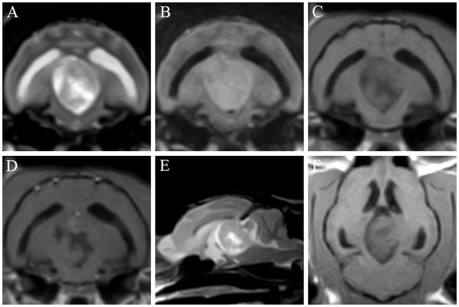
A 15.5-year-old, neutered, male, domestic shorthair cat was presented with neurologic dysfunctions. At presentation, an obtunded mental status and vestibular ataxia were identified. On neurologic examination, postural reactions were decreased-to-absent in all four limbs, and pupillary light reflexes showed bilaterally delayed results. Magnetic resonance imaging was performed, and a demarcated lesion was identified in the third ventricle. The cat was tentatively diagnosed with a brain tumor, which was suspected to be a meningioma. The cat was treated with hydroxyurea and prednisolone. Mental status was considered more alert, and ataxia improved following treatment. On the 106th day after the commencement of treatment, a 18F-fluorodeoxyglucose (FDG)-positron emission tomography (PET) scan was performed. On the PET images, a hypermetabolic region was found in the lesion. The average standardized uptake value of FDG was 2.47, and the tumor-to-normal-tissue ratio was 1.25. The cat died 408 days following the commencement of treatment, and a grade 1 meningioma was confirmed by postmortem histopathology. Immunohistochemistry for Ki-67 and p53 was performed. The labeling indices of Ki-67 and p53 were 2.56 and 0%, respectively. This case shows that chemotherapy with hydroxyurea and prednisolone may be considered in the treatment of feline meningiomas. Furthermore, this is the first case describing the application of FDG-PET to visualize a naturally occurring meningioma in a cat.
Keywords: brain tumor; chemotherapy; feline; hydroxyurea; positron emission tomography.
Copyright © 2021 Yun, Koo, Kim, Lee, Kim, Jung, Yang and Kang.
Conflict of interest statement
The authors declare that the research was conducted in the absence of any commercial or financial relationships that could be construed as a potential conflict of interest.
Figures


Similar articles
-
Characteristics of 18F-FDG and 18F-FDOPA PET in an 8-year-old neutered male Yorkshire Terrier dog with glioma: long-term chemotherapy using hydroxyurea plus imatinib with prednisolone and immunoreactivity for PDGFR-β and LAT1.Vet Q. 2021 Dec;41(1):163-171. doi: 10.1080/01652176.2021.1906466. Vet Q. 2021. PMID: 33745419 Free PMC article.
-
Case Report: 18F-FDOPA PET in the clinical management of a dog with an intraventricular tumor suspected to be choroid plexus papilloma.Front Vet Sci. 2025 Mar 26;12:1477063. doi: 10.3389/fvets.2025.1477063. eCollection 2025. Front Vet Sci. 2025. PMID: 40206257 Free PMC article.
-
Case Report: 18F-Fluoro-L-Phenylalanine Positron Emission Tomography Findings and Immunoreactivity for L-Type Amino Acid Transporter 1 in a Dog With Meningioma.Front Vet Sci. 2022 Jul 15;9:899229. doi: 10.3389/fvets.2022.899229. eCollection 2022. Front Vet Sci. 2022. PMID: 35909694 Free PMC article.
-
1-11C-acetate versus 18F-FDG PET in detection of meningioma and monitoring the effect of gamma-knife radiosurgery.J Nucl Med. 2010 Jun;51(6):883-91. doi: 10.2967/jnumed.109.070565. Epub 2010 May 19. J Nucl Med. 2010. PMID: 20484430
-
Grade I lymphomatoid granulomatosis with increased uptake of [18F] fluorodeoxyglucose in positron emission tomography: a case report.J Clin Exp Hematop. 2009 May;49(1):39-44. doi: 10.3960/jslrt.49.39. J Clin Exp Hematop. 2009. PMID: 19474516 Review.
Cited by
-
Domestic Animal Models of Central Nervous System Tumors: Focus on Meningiomas.Life (Basel). 2023 Nov 30;13(12):2284. doi: 10.3390/life13122284. Life (Basel). 2023. PMID: 38137885 Free PMC article. Review.
-
Surgical treatment of feline intracranial meningiomas: a retrospective study of 26 cases.J Vet Sci. 2024 Mar;25(2):e25. doi: 10.4142/jvs.23207. J Vet Sci. 2024. PMID: 38568826 Free PMC article.
-
Case report: Evaluation of hindlimb ischemia using 18F-fluorodeoxyglucose positron emission tomography in a cat with cardiogenic arterial thromboembolism.Front Vet Sci. 2023 Sep 6;10:1223866. doi: 10.3389/fvets.2023.1223866. eCollection 2023. Front Vet Sci. 2023. PMID: 37745211 Free PMC article.
References
Publication types
LinkOut - more resources
Full Text Sources
Other Literature Sources
Research Materials
Miscellaneous