

Acute hemorrhagic leukoencephalitis in a COVID-19 patient-a case report with literature review
- PMID: 33575849
- PMCID: PMC7878029
- DOI: 10.1007/s00234-021-02667-1
Acute hemorrhagic leukoencephalitis in a COVID-19 patient-a case report with literature review
Abstract
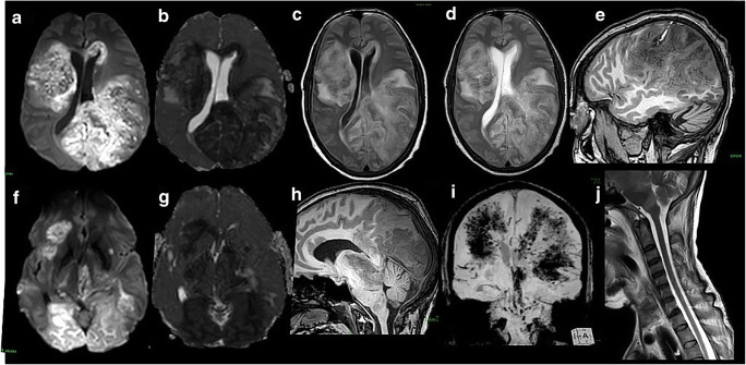
Purpose: Acute hemorrhagic leukoencephalitis (AHLE) is a rare and severe form of acute disseminated encephalomyelitis (ADEM). Only a few reports of AHLE in coronavirus disease 2019 (COVID-19) patients have been described to date. We report a case of COVID-19-related AHLE along with a literature review describing salient clinical and imaging characteristics.
Methods: A literature search was performed on Medline (2020-present), PubMed, Cochrane Library, CINAHL, and Google scholar on 28 January 2021 for all articles published using MeSH terms "COVID-19" or "SARS-CoV-2" with "Acute hemorrhagic leukoencephalitis" or "Acute hemorrhagic encephalitis." Relevant case reports and case series describing clinical and imaging features of AHLE associated with SARS-CoV-2 infection were included, data compiled, and critically reviewed.
Results: Acute onset encephalopathy and rapidly deteriorating neurological status is the common clinical presentation in AHLE. CSF analysis reveals elevated proteins and lymphocytic pleocytosis. Typical neuroimaging features include multifocal, variable-sized, poorly defined cerebral white matter lesions with cortical sparing. Involvement of the brainstem, cerebellar peduncles, and deep grey matter can also occur, although rarely. Lesions are hyperintense on T2-weighted (T2W) and fluid-attenuated inversion recovery (FLAIR) images, hypointense on T1W images, and show microhemorrhages, variable diffusion restriction, and post-contrast enhancement. Extensive microhemorrhages, brainstem involvement, and gross hemorrhage often portend a poor prognosis.
Conclusion: Heightened awareness about the clinical and imaging presentation of COVID-19-related AHLE can positively alter the outcome in a select few by enabling early diagnosis and aggressive management.
Keywords: Acute disseminated encephalomyelitis (ADEM); Acute hemorrhagic leukoencephalitis (AHLE); COVID-19; Computed tomography (CT); Magnetic resonance imaging (MRI).
Conflict of interest statement
The authors declare that they have no conflict of interest/competing interests.
Figures


References
-
- Bhatt P, Bray L, Raju S, Dapaah-Siakwan F, Patel A, Chaudhari R, et al. Temporal trends of pediatric hospitalizations with acute disseminated encephalomyelitis in the United States: An Analysis from 2006 to 2014 using National Inpatient Sample. J Pediatr. 2019;206:26–32.e1. doi: 10.1016/j.jpeds.2018.10.044. - DOI - PubMed
Publication types
MeSH terms
LinkOut - more resources
Full Text Sources
Other Literature Sources
Medical
Miscellaneous
