. 2021 May;31(3):e12942.
doi: 10.1111/bpa.12942. Epub 2021 Feb 12.

Cognitive decline in amyotrophic lateral sclerosis: Neuropathological substrate and genetic determinants

Affiliations

Cognitive decline in amyotrophic lateral sclerosis: Neuropathological substrate and genetic determinants

Sergi Borrego-Écija et al. Brain Pathol. 2021 May.

Abstract

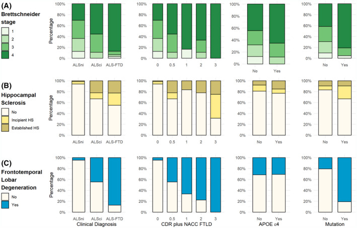
Cognitive impairment and behavioral changes in amyotrophic lateral sclerosis (ALS) are now recognized as part of the disease. Whether it is solely related to the extent of TDP-43 pathology is currently unclear. We aim to evaluate the influence of age, genetics, neuropathological features, and concomitant pathologies on cognitive impairment in ALS patients. We analyzed a postmortem series of 104 ALS patients and retrospectively reviewed clinical and neuropathological data. We assessed the burden and extent of concomitant pathologies, the role of APOE ε4 and mutations, and correlated these findings with cognitive status. We performed a logistic regression model to identify which pathologies are related to cognitive impairment. Cognitive decline was recorded in 38.5% of the subjects. Neuropathological features of frontotemporal lobar degeneration (FTLD) were found in 32.7%, explaining most, but not all, cases with cognitive impairment. Extent of TDP-43 pathology and the presence of hippocampal sclerosis were associated with cognitive impairment. Mutation carriers presented a higher burden of TDP-43 pathology and FTLD more frequently than sporadic cases. Most cases (89.4%) presented some degree of concomitant pathologies. The presence of concomitant pathologies was associated with older age at death. FTLD, but also Alzheimer's disease, were the predominant underlying pathologies explaining the cognitive impairment in ALS patients. In sum, FTLD explained the presence of cognitive decline in most but not all ALS cases, while other non-FTLD related findings can influence the cognitive status, particularly in older age groups.

Keywords: ALS-FTD; Alzheimer’s disease; TDP-43 protein; amyotrophic lateral sclerosis; frontotemporal dementia; neuropathology.


Conflict of interest statement

The authors declare that they have no conflict of interest.

Figures

FIGURE 1
FIGURE 1
Characteristic neuropathological features of ALS. (A) Cross section through the thoracic spinal cord shows prominent degeneration of the lateral and anterior corticospinal tract and atrophy of the anterior horns (higher magnification in E). There is also myelin pallor in anterior roots compared to posterior roots. The degeneration of the lateral corticospinal tract is characterized by loss of myelin sheaths (B), reduction of axonal profiles (C), and increased macrophagic activity (D), as compared with posterior horns (b, c, d). (F) some residual motor neurons (here from the n. XII) may contain granular eosinophilic cytoplasmic inclusions or Bunina bodies (arrow and inset). Others may appear as pale spherical inclusions (G). Axonal damage with swellings (H) in anterior horns is well depicted by anti‐neurofilament immunohistochemistry (large brown structures). I–L: Immunohistochemistry for pTDP‐43 reveals a spectrum of inclusions including a fine granular pattern in large motor neurons (I), fibrillar and skein like inclusions (J), compact spherical neuronal inclusions (K), and coiled‐body like inclusions in oligodendrocytes (L). Scale bars: 20 µm in Bb, Cc, Dd, F; 10 µm in G–L, 100 µm in E
FIGURE 2
FIGURE 2
Barplots representing the proportion of ALS cases with (A) the different Brettschneider stage, (B) the presence of hippocampal sclerosis and (C) Frontotemporal Lobar degeneration according to their clinical diagnosis, CDR plus NACC FTLD stage, presence of APOE ε4, or presence of mutations in genes reported as causative of or at risk for ALS/FTLD
FIGURE 3
FIGURE 3
Frequent co‐pathologies in ALS. (A–C) Segmental hippocampal sclerosis is sometimes better identified in more anterior segments of the hippocampus (A, arrowheads). It is characterized by neuronal loss and prominent fibrillar gliosis (B) in the CA1 sector and subiculum. In most cases granule cells of the dentate gyrus harbor TDP‐43 protein inclusions in the cytoplasm (C, upper row) and in some cases there are abundant fine threads in the CA1 sector within and adjacent to the sclerotic area (C, lower row). (D and E) Argyrophilic grain pathology is well depicted by the AT8 anti‐tau antibody (D) and consists of grain‐like structures along neuronal processes, abundant threads, and a diffuse cytoplasmic neuronal staining (pretangle type) instead of compact tangles. They are frequently associated with ballooned neurons in the amygdala (E, upper row) and oligodendroglial coiled bodies in temporomedial white matter (E, lower row). Astrocytic tau pathology in form of granular fuzzy astrocytes (F, upper row) and thorn‐shaped astrocytes in the glia limitans (F, lower row, here as subependymal and perivascular thorn‐shaped astrocyte) are features of age‐related tau astrogliopathy (ARTAG). (G and H) Alzheimer’s disease neuropathologic changes have been also found as co‐pathology in a fraction of cases. It is characterized by dense cored amyloid plaques with or without amyloid angiopathy (G) and tau positive neurofibrillary pathology with tangles, neuropil threads, and dystrophic neurites around amyloid deposits (H). (I) Lewy body pathology has been detected less frequently and can be encountered in brainstem neurons (HE, upper row, medulla oblongata) and/or limbic system and is well identified with anti‐alpha‐synuclein antibodies (lower row), where Lewy‐neurites are also frequently seen (here in the locus coeruleus). Scale bars: 50 µm in B, F, G, H; 20 µm in C (upper and lower panel), D, E, and I (upper and lower panel)
FIGURE 4
FIGURE 4
Presence of total concomitant pathologies in ALS cases (A), distribution of pathology according to age at death (B), distribution of pathology according to the CDR NACC plus FTLD staging (C), and distribution of pathology according to the presence of APOE ε4 allele (D). Neurofibrillary tangles were staged following Braak stages (24). Amyloid represents Aβ Thal phases (25). Globally, Alzheimer’s disease pathology was staged according the NIA/AA guidelines and an ABC score was assigned (27). Cerebral Amyloid angiopathy (CAA) has been reported as absent or present. Vascular lesions were classified according to small or large vessel pathology. Assignments of Lewy Body pathology were performed following McKeith criteria (30). Possible or definite primary age related tauopathy (PART) was assessed according the current neuropathological criteria (28). Age‐related tau astrogliopathy (ARTAG) was evaluated according the current criteria (29). Argyrophilic grain disease (AGD) was staged according to the Saito criteria (31). Granulovacuolar degeneration (GVD) was staged according to Thal et al (33)

References

    1. Goldstein LH, Abrahams S. Changes in cognition and behaviour in amyotrophic lateral sclerosis: nature of impairment and implications for assessment. Lancet Neurol. 2013;12:368–80. - PubMed
    1. Strong MJ, Abrahams S, Goldstein LH, Woolley S, Mclaughlin P, Snowden J, et al. Amyotrophic lateral sclerosis ‐ frontotemporal spectrum disorder (ALS‐FTSD): revised diagnostic criteria. Amyotroph Lateral Scler Front Degener. 2017;18:153–74. - PMC - PubMed
    1. Prudlo J, König J, Schuster C, Kasper E, Büttner A, Teipel S, et al. TDP‐43 pathology and cognition in ALS. Neurology. 2016;87:1019–23. - PubMed
    1. Saxon JA, Harris JM, Thompson JC, Jones M, Richardson AMT, Langheinrich T, et al. Semantic dementia, progressive non‐fluent aphasia and their association with amyotrophic lateral sclerosis. J Neurol Neurosurg Psychiatry. 2017;88:711–12. - PMC - PubMed
    1. Strong MJ, Grace GM, Freedman M, Lomen‐Hoerth C, Woolley S, Goldstein LH, et al. Consensus criteria for the diagnosis of frontotemporal cognitive and behavioural syndromes in amyotrophic lateral sclerosis. Amyotroph Lateral Scler. 2009;10:131–46. - PubMed

Publication types

MeSH terms