

Application of laser capture microdissection and PCR sequencing in the diagnosis of Coccidioides spp. infection: A case report and literature review in China
- PMID: 33576325
- PMCID: PMC7919914
- DOI: 10.1080/22221751.2021.1889931
Application of laser capture microdissection and PCR sequencing in the diagnosis of Coccidioides spp. infection: A case report and literature review in China
Abstract
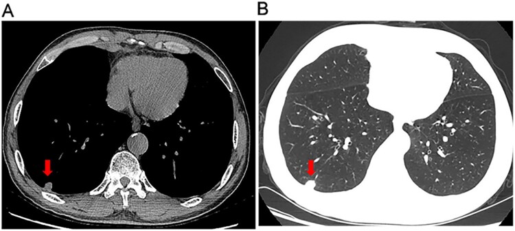
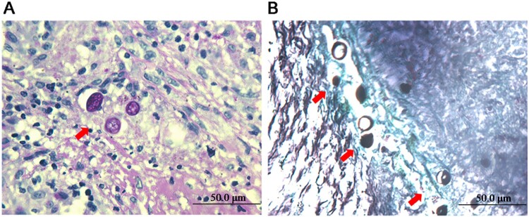
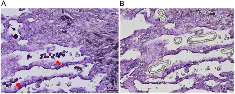
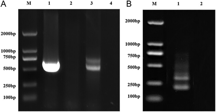
Coccidioidomycosis is endemic to California, Arizona, and Mexico. In recent years, the reported cases of coccidioidomycosis have increased in nonendemic regions. Here, we reported a case of imported pulmonary coccidioidomycosis in a Chinese patient. A 63-year-old man presented with dry cough and fatigue for 6 months, and a computed tomography scan revealed a solitary nodule in the right lower lung and small nodules in both lungs. The diagnosis of coccidioidomycosis was initially confirmed by histopathologic examination. The pathogen Coccidioides spp. was identified by laser capture microdissection (LCM) combined with subsequent molecular techniques based on the positive histopathologic features. Additionally, we reviewed 47 reported cases of coccidioidomycosis in China. The number of reported cases is increasing, and the incidence of disseminated infection has exhibited a trend of shifting towards healthy young adults in China. Since clinical presentations and imaging findings lack specificity, a majority of domestic cases of coccidioidomycosis were initially misdiagnosed as tumours or tuberculosis. Moreover, the diagnosis of endemic mycoses may be challenging because of their rarity and the limited availability of diagnostic tests. The diagnosis was mainly confirmed by histopathological examination. The species involved were identified based on positive cultures in only 4 cases. To our knowledge, this is the first study to use LCM and molecular techniques to identify Coccidioides spp. in the histopathologically positive but uncultivable specimen. Comparing with previous reported studies, LCM combined with nucleic acid amplification techniques improve the ability of species identification for the timely diagnosis of coccidioidomycosis.
Keywords: Coccidioides spp; Coccidioidomycosis; laser capture microdissection; molecular diagnosis.
Conflict of interest statement
No potential conflict of interest was reported by the author(s).
Figures
Similar articles
-
Coccidioidomycosis masquerading as skeletal tuberculosis: an imported case and review of coccidioidomycosis in India.Trop Doct. 2014 Jan;44(1):25-8. doi: 10.1177/0049475513512641. Epub 2013 Nov 21. Trop Doct. 2014. PMID: 24265192 Review.
-
[A case of coccidioidomycosis in Turkey imported from the United States of America].Mikrobiyol Bul. 2017 Apr;51(2):183-190. doi: 10.5578/mb.54033. Mikrobiyol Bul. 2017. PMID: 28566083 Turkish.
-
Molecular markers in the epidemiology and diagnosis of coccidioidomycosis.Rev Iberoam Micol. 2014 Jan-Mar;31(1):49-53. doi: 10.1016/j.riam.2013.09.011. Epub 2013 Nov 20. Rev Iberoam Micol. 2014. PMID: 24270069 Review.
-
A case study of imported pulmonary coccidioidomycosis.J Zhejiang Univ Sci B. 2011 Apr;12(4):298-302. doi: 10.1631/jzus.B1000261. J Zhejiang Univ Sci B. 2011. PMID: 21462386 Free PMC article.
-
Coccidioidomycosis: Imported and possible domestic cases in China: A case report and review, 1958-2017.Mycoses. 2018 Jul;61(7):506-513. doi: 10.1111/myc.12750. Epub 2018 Feb 19. Mycoses. 2018. PMID: 29383771 Review.
Cited by
-
The first suspected disseminated Hormographiella aspergillata infection in China, diagnosed using metagenomic next-generation sequencing: a case report and literature review.Emerg Microbes Infect. 2023 Dec;12(1):2220581. doi: 10.1080/22221751.2023.2220581. Emerg Microbes Infect. 2023. PMID: 37254739 Free PMC article.
-
Laser Capture Proteomics: spatial tissue molecular profiling from the bench to personalized medicine.Expert Rev Proteomics. 2021 Oct;18(10):845-861. doi: 10.1080/14789450.2021.1984886. Epub 2021 Dec 14. Expert Rev Proteomics. 2021. PMID: 34607525 Free PMC article. Review.
-
Combination of metagenomic next-generation sequencing and morphology for identifying Coccidioides immitis: a case report.Front Med (Lausanne). 2025 Jan 22;11:1500014. doi: 10.3389/fmed.2024.1500014. eCollection 2024. Front Med (Lausanne). 2025. PMID: 39911666 Free PMC article.
-
Advances in single-cell sequencing technology in microbiome research.Genes Dis. 2023 Sep 28;11(4):101129. doi: 10.1016/j.gendis.2023.101129. eCollection 2024 Jul. Genes Dis. 2023. PMID: 38545125 Free PMC article. Review.
-
Pulmonary coccidioidomycosis in China: Case reports and literature review.IDCases. 2024 Oct 18;38:e02102. doi: 10.1016/j.idcr.2024.e02102. eCollection 2024. IDCases. 2024. PMID: 39507637 Free PMC article.
References
-
- Stockamp NW. Thompson GR 3rd. coccidioidomycosis. Infect Dis Clin North Am. 2016;30(1):229–246. - PubMed
-
- Liang G, Shen Y, Lv G, et al. . Coccidioidomycosis: imported and possible domestic cases in China: A case report and review, 1958-2017. Mycoses. 2018;61(7):506–513. - PubMed
-
- Malo J, Luraschi-Monjagatta C, Wolk DM, et al. . Update on the diagnosis of pulmonary coccidioidomycosis. Ann Am Thorac Soc. 2014;11(2):243–253. - PubMed
-
- Canteros CE, Vélez HA, Toranzo AI, et al. . Molecular identification of Coccidioides immitis in formalin-fixed, paraffin-embedded (FFPE) tissues from a Colombian patient. Med Mycol. 2015;53(5):520–527. - PubMed
-
- Sheff KW, York ER, Driebe EM, et al. . Development of a rapid, cost-effective TaqMan real-time PCR assay for identification and differentiation of Coccidioides immitis and Coccidioides posadasii. Med Mycol. 2010;48(3):466–469. - PubMed
Publication types
MeSH terms
LinkOut - more resources
Full Text Sources
Other Literature Sources
Medical
Miscellaneous