

The effects of cardiac stretch on atrial fibroblasts: analysis of the evidence and potential role in atrial fibrillation
- PMID: 33576384
- PMCID: PMC8803074
- DOI: 10.1093/cvr/cvab035
The effects of cardiac stretch on atrial fibroblasts: analysis of the evidence and potential role in atrial fibrillation
Abstract
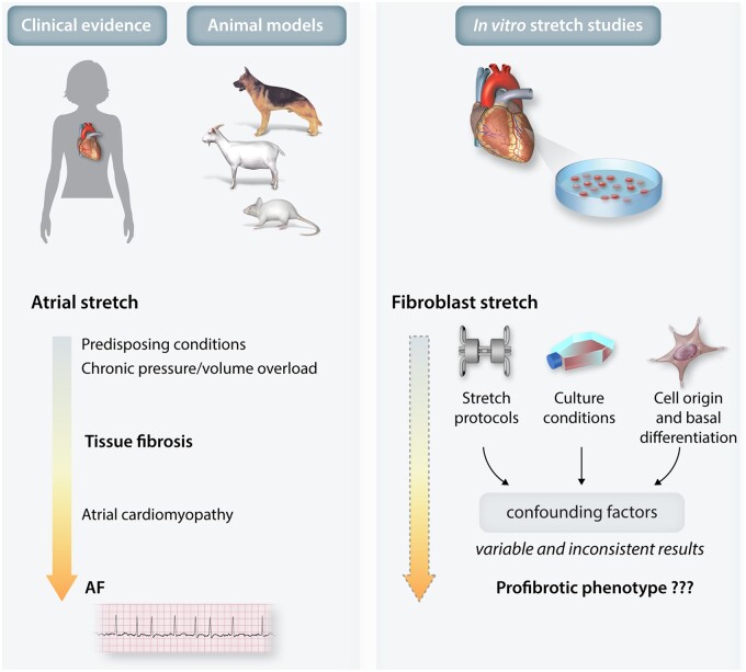
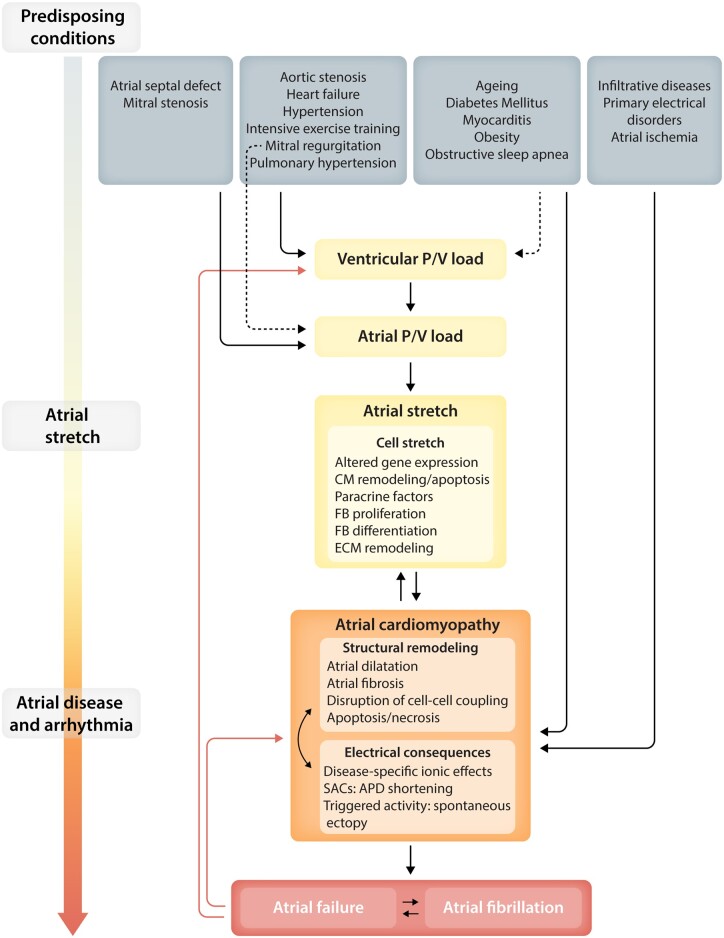
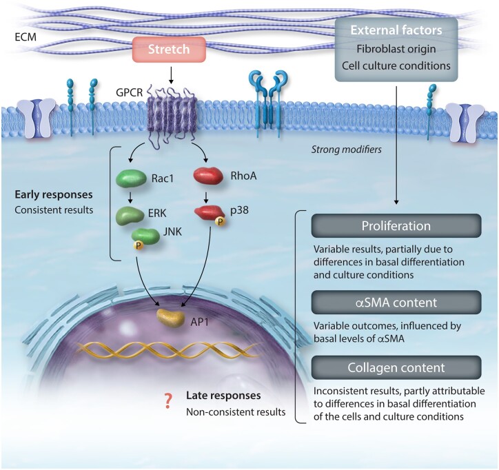
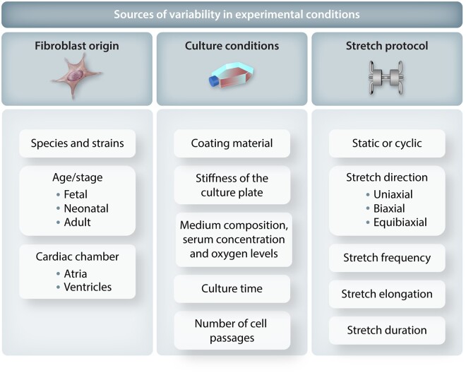
Atrial fibrillation (AF) is an important clinical problem. Chronic pressure/volume overload of the atria promotes AF, particularly via enhanced extracellular matrix (ECM) accumulation manifested as tissue fibrosis. Loading of cardiac cells causes cell stretch that is generally considered to promote fibrosis by directly activating fibroblasts, the key cell type responsible for ECM production. The primary purpose of this article is to review the evidence regarding direct effects of stretch on cardiac fibroblasts, specifically: (i) the similarities and differences among studies in observed effects of stretch on cardiac fibroblast function; (ii) the signalling pathways implicated; and (iii) the factors that affect stretch-related phenotypes. Our review summarizes the most important findings and limitations in this area and gives an overview of clinical data and animal models related to cardiac stretch, with particular emphasis on the atria. We suggest that the evidence regarding direct fibroblast activation by stretch is weak and inconsistent, in part because of variability among studies in key experimental conditions that govern the results. Further work is needed to clarify whether, in fact, stretch induces direct activation of cardiac fibroblasts and if so, to elucidate the determining factors to ensure reproducible results. If mechanical load on fibroblasts proves not to be clearly profibrotic by direct actions, other mechanisms like paracrine influences, the effects of systemic mediators and/or the direct consequences of myocardial injury or death, might account for the link between cardiac stretch and fibrosis. Clarity in this area is needed to improve our understanding of AF pathophysiology and assist in therapeutic development.
Keywords: Atrial fibrillation; Cardiac fibroblast; Fibrosis; Mechanical strain; Pressure overload; Stretch.
Published on behalf of the European Society of Cardiology. All rights reserved. © The Author(s) 2021. For permissions, please email: journals.permissions@oup.com.
Figures
Similar articles
-
Heterogeneous atrial wall thickness and stretch promote scroll waves anchoring during atrial fibrillation.Cardiovasc Res. 2012 Apr 1;94(1):48-57. doi: 10.1093/cvr/cvr357. Epub 2012 Jan 6. Cardiovasc Res. 2012. PMID: 22227155 Free PMC article.
-
Caveolin-1 Deficiency Induces Atrial Fibrosis and Increases Susceptibility to Atrial Fibrillation by the STAT3 Signaling Pathway.J Cardiovasc Pharmacol. 2021 Aug 1;78(2):175-183. doi: 10.1097/FJC.0000000000001066. J Cardiovasc Pharmacol. 2021. PMID: 34554674
-
Matrine reduces susceptibility to postinfarct atrial fibrillation in rats due to antifibrotic properties.J Cardiovasc Electrophysiol. 2018 Apr;29(4):616-627. doi: 10.1111/jce.13448. Epub 2018 Feb 27. J Cardiovasc Electrophysiol. 2018. PMID: 29377366
-
Interleukin-6: Molecular Mechanisms and Therapeutic Perspectives in Atrial Fibrillation.Curr Med Sci. 2025 Apr;45(2):157-168. doi: 10.1007/s11596-025-00021-7. Epub 2025 Mar 4. Curr Med Sci. 2025. PMID: 40035997 Review.
-
Molecular and Cellular Mechanisms of Atrial Fibrosis in Atrial Fibrillation.JACC Clin Electrophysiol. 2017 May;3(5):425-435. doi: 10.1016/j.jacep.2017.03.002. Epub 2017 May 15. JACC Clin Electrophysiol. 2017. PMID: 29759598 Review.
Cited by
-
Atrial fibrillation episode status and incidence of coronary slow flow: A propensity score-matched analysis.Front Cardiovasc Med. 2023 Mar 20;10:1047748. doi: 10.3389/fcvm.2023.1047748. eCollection 2023. Front Cardiovasc Med. 2023. PMID: 37020520 Free PMC article.
-
Atrial fibrillation variant-to-gene prioritization through cross-ancestry eQTL and single-nucleus multiomic analyses.iScience. 2024 Aug 5;27(9):110660. doi: 10.1016/j.isci.2024.110660. eCollection 2024 Sep 20. iScience. 2024. PMID: 39262787 Free PMC article.
-
Cardiac remodeling in patients with atrial fibrillation reversing bradycardia-induced cardiomyopathy: A case report.World J Clin Cases. 2024 Mar 6;12(7):1339-1345. doi: 10.12998/wjcc.v12.i7.1339. World J Clin Cases. 2024. PMID: 38524509 Free PMC article.
-
Translation of pathophysiological mechanisms of atrial fibrosis into new diagnostic and therapeutic approaches.Nat Rev Cardiol. 2025 Apr;22(4):225-240. doi: 10.1038/s41569-024-01088-w. Epub 2024 Oct 23. Nat Rev Cardiol. 2025. PMID: 39443702 Review.
-
Cardiac fibroblasts and mechanosensation in heart development, health and disease.Nat Rev Cardiol. 2023 May;20(5):309-324. doi: 10.1038/s41569-022-00799-2. Epub 2022 Nov 14. Nat Rev Cardiol. 2023. PMID: 36376437 Review.
References
-
- Andrade J, Khairy P, Dobrev D, Nattel S.. The clinical profile and pathophysiology of atrial fibrillation: relationships among clinical features, epidemiology, and mechanisms. Circ Res 2014;114:1453–1468. - PubMed
-
- Satoh T, Zipes DP.. Unequal atrial stretch in dogs increases dispersion of refractoriness conducive to developing atrial fibrillation. J Cardiovasc Electrophysiol 1996;7:833–842. - PubMed
-
- Sideris DA, Toumanidis ST, Thodorakis M, Kostopoulos K, Tselepatiotis E, Langoura C, Stringli T, Moulopoulos SD.. Some observations on the mechanism of pressure related atrial fibrillation. Eur Heart J 1994;15:1585–1589. - PubMed
-
- Gottdiener JS, Seliger S, deFilippi C, Christenson R, Baldridge AS, Kizer JR, Psaty BM, Shah SJ.. Relation of biomarkers of cardiac injury, stress, and fibrosis with cardiac mechanics in patients ≥ 65 years of age. Am J Cardiol Elsevier 2020;136:156–163. - PubMed
Publication types
MeSH terms
LinkOut - more resources
Full Text Sources
Other Literature Sources
Medical
