Review
. 2021 Sep 28;117(11):2326-2339.
doi: 10.1093/cvr/cvab046.

Vascular smooth muscle cells in atherosclerosis: time for a re-assessment

Affiliations
Review

Vascular smooth muscle cells in atherosclerosis: time for a re-assessment

Mandy O J Grootaert et al. Cardiovasc Res. .

Abstract

Vascular smooth muscle cells (VSMCs) are key participants in both early and late-stage atherosclerosis. VSMCs invade the early atherosclerotic lesion from the media, expanding lesions, but also forming a protective fibrous cap rich in extracellular matrix to cover the 'necrotic' core. Hence, VSMCs have been viewed as plaque-stabilizing, and decreased VSMC plaque content-often measured by expression of contractile markers-associated with increased plaque vulnerability. However, the emergence of lineage-tracing and transcriptomic studies has demonstrated that VSMCs comprise a much larger proportion of atherosclerotic plaques than originally thought, demonstrate multiple different phenotypes in vivo, and have roles that might be detrimental. VSMCs down-regulate contractile markers during atherosclerosis whilst adopting alternative phenotypes, including macrophage-like, foam cell-like, osteochondrogenic-like, myofibroblast-like, and mesenchymal stem cell-like. VSMC phenotypic switching can be studied in tissue culture, but also now in the media, fibrous cap and deep-core region, and markedly affects plaque formation and markers of stability. In this review, we describe the different VSMC plaque phenotypes and their presumed cellular and paracrine functions, the regulatory mechanisms that control VSMC plasticity, and their impact on atherogenesis and plaque stability.

Keywords: Atherosclerosis; Vascular smooth muscle.


Figures

Figure 1
Figure 1
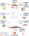
Regulation and characterisation of VSMC phenotypic switching. In the healthy vessel wall, VSMCs exhibit a contractile phenotype characterized by expression of contractile proteins (ACTA2, TAGLN, and MYH11) regulated by MYOCD, which is negatively regulated by NFκB, FOXO3A, miRNA221/222, miRNA21, and miRNA124. Upon injury or atherosclerosis, VSMCs switch to a synthetic phenotype mediated via PDGF-BB and Klf4, which is inhibited by miRNA143/145 and TGFβ. Synthetic VSMCs are characterised by increased secretion of ECM, MMPs, pro-inflammatory cytokines, and exosomes. Exosomes can trigger neighbouring VSMCs to transdifferentiate into osteochondrogenic VSMCs characterised by RUNX2, SOX9, osteopontin expression, release of calcium deposits, and calcifying vesicles. Osteochondrocyte-like VSMCs secrete calcifying vesicles that further propagate calcification. In some cases, calcifying VSMCs originate from medial contractile VSMCs in response to high concentrations of calcium and phosphate, β-catenin/Wnt signalling, KLF4, or miRNA221/222. Exposure to agLDL or oxLDL triggers a switch to macrophage-like VSMCs or foam cells characterised by CD68, CD36, LGALS3, CD11b, and pro-inflammatory cytokine expression, which is inhibited by integrin β3, apolipoprotein A1, or HDL. VSMCs may adopt adipocyte-like features in a corticosteroid/insulin-rich milieu characterised by expression of adipsin, leptin, and PPARγ pathway activation. KLF4 promotes phenotypic modulation into macrophage-like and Sca1-expressing mesenchymal-like VSMCs, while MSCs differentiate into VSMCs in vitro when exposed to TGFβ and PDGF-BB. VSMCs might also derive from ECs following EndMT driven by TGFβ signalling. VSMCs can potentially transdifferentiate into endothelial-like cells characterised by CD31 expression and angiogenic features in response to shear stress or via a KLF4-dependent stem cell state. TCF21 and OCT4 promote phenotypic modulation into an athero-protective ‘myofibroblast’-like VSMC characterised by fibronectin, collagen 1 alpha 1, and proteoglycans production. Phenotypically switched VSMCs conserve an epigenetic H3K4me2 signature of their former contractile state that can be used as a VSMC-specific marker. MYOCD, myocardin; NFκB, nuclear factor kappa-light-chain-enhancer of activated B cells; FOXO3a, forkhead box O3a; PDGF-BB, platelet-derived growth factor; KLF4, Krüppel-like factor 4; ECM, extracellular matrix; MMP, matrix metalloproteinases; RUNX2, runt-related transcription factor 2; SOX9, SRY-related HMG-box 9; PPARγ, peroxisome proliferator-activated receptors gamma; Sca1, stem cell antigen 1; TGFβ, transforming growth factor beta; MSCs, mesenchymal stem cells; EndMT, endothelial-to-mesenchymal transition; TCF21, transcription factor 21; OCT4, octamer binding transcription factor 4.
Figure 2
Figure 2
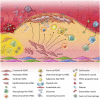
The role of VSMCs in the pathogenesis of atherosclerosis. Schematic of all VSMCs phenotypes found in mouse and/or human atherosclerotic plaques to date including their location within the lesion, although their presence and/or abundance may vary depending on the lesion stage. The medial layer is composed of different VSMC clones (illustrated as red, blue, purple, and green) exhibiting a contractile phenotype. During atherogenesis, medial VSMCs underlying the lesion lose their contractile markers and undergo phenotypic switching, likely upon exposure to lipids or calcification-promoting molecules. Only one or a small number of phenotypically modulated medial VSMC clones (here in red) invade and occupy the lesion. The mechanism for oligoclonal expansion is still unclear but may involve a subpopulation of Sca1+ VSMCs that are predisposed to invade the lesion or express Sca1 during clonal expansion. Within the lesion, these modulated VSMCs adopt various phenotypes: synthetic, macrophage-like, foam cells, MSC-like, osteochondrogenic, myofibroblast-like/fibromyocyte and potentially adipocyte-like, and EC-like VSMCs. Synthetic and myofibroblast-like/fibromyocyte VSMC clones occupy the fibrous cap and produce ECM. Macrophage-like and foam cell-like VSMCs possess requisite markers but still differ from professional monocytes that enter the lesion via the circulation. In response to various cues, VSMC undergoes apoptosis characterised by formation of apoptotic bodies and/or undergo (secondary) necrosis and accumulate in the cholesterol-rich ‘necrotic’ core. Osteochondrogenic VSMCs promote micro- and macro-calcification that accumulate in the fibrous cap and necrotic core, respectively. Calcifying and synthetic VSMCs secrete EVs or exosomes carrying various cargos (e.g. nucleic acids and bioactive molecules) that are released into the circulation upon plaque rupture and can potentially be used as CVD biomarkers. Sca1, stem cell antigen 1; ECM, extracellular matrix.
Figure 3
Figure 3
Regulatory pathways and putative role of different VSMC phenotypes on plaque stability. The major regulatory pathways controlling the transition from contractile to phenotypically modified VSMCs include the pluripotency factors KLF4 and OCT4, growth factors PDGF-BB and TGFβ, and NFκB. KLF4 represses expression of contractile genes via multiple mechanisms including binding to SRF, thereby antagonizing MYOCD tethering to SRF. OCT4 is transcriptionally regulated by KLF4 but plays an opposite role in VSMC phenotypic switching. NFκB decreases MYOCD expression by binding to its promoter leading to reduced expression of contractile genes. PDGF-BB suppresses contractile gene expression via different mechanisms, which partially involves KLF4. The contractile phenotype is positively controlled by the growth factor TGFβ and miRNAs miR143 and miR145 that work partially in concert to down-regulate klf4. Also epigenetic regulation by histone acetylation (Ac) and methylation (Me) at the promoter of contractile genes facilitates VSMC differentiation by increasing chromatin accessibility for MYOCD/SRF complexes. Phenotypic switching results in a range of different VSMC phenotypes, some of which are postulated to have predominantly negative effects (foam cell and macrophage-like), neutral effects (osteochondrogenic or synthetic VSMCs), or mostly positive effects on plaque stability (mesenchymal cell- or myofibroblast-like/fibromyocyte cells). Solid evidence for EC-like and adipocyte-like VSMCs in atherosclerosis is currently lacking. To date, only myofibroblast-like VSMCs have been truly associated with plaque stability in mice, although the ability of VSMCs to undergo reprogramming into a stem cell-like state has potential to transdifferentiate into athero-protective VSMC subtypes. TGFβ, transforming growth factor beta; PDGF-BB, platelet-derived growth factor; KLF4, Krüppel-like factor 4; OCT4, octamer binding transcription factor; SRF, serum response factor; MYOCD, myocardin.

References

    1. Yahagi K, Kolodgie FD, Otsuka F, Finn AV, Davis HR, Joner M, Virmani R.. Pathophysiology of native coronary, vein graft, and in-stent atherosclerosis. Nat Rev Cardiol 2016;13:79–98. - PubMed
    1. Barquera S, Pedroza-Tobias A, Medina C, Hernandez-Barrera L, Bibbins-Domingo K, Lozano R, Moran AE.. Global overview of the epidemiology of atherosclerotic cardiovascular disease.Arch Med Res 2015;46:328–338. - PubMed
    1. Franck G, Mawson T, Sausen G, Salinas M, Masson GS, Cole A, Beltrami-Moreira M, Chatzizisis Y, Quillard T, Tesmenitsky Y, Shvartz E, Sukhova GK, Swirski FK, Nahrendorf M, Aikawa E, Croce KJ, Libby P.. Flow perturbation mediates neutrophil recruitment and potentiates endothelial injury via TLR2 in mice: implications for superficial erosion. Circ Res 2017;121:31–42. - PMC - PubMed
    1. Kolodgie FD, Burke AP, Farb A, Weber DK, Kutys R, Wight TN, Virmani R.. Differential accumulation of proteoglycans and hyaluronan in culprit lesions: insights into plaque erosion. ATVB 2002;22:1642–1648. - PubMed
    1. Owens GK.Regulation of differentiation of vascular smooth muscle cells.Physiol Rev 1995;75:487–517. - PubMed

Publication types