. 2021 Apr;47(4):48.
doi: 10.3892/ijmm.2021.4881. Epub 2021 Feb 12.

Apoptotic mechanism activated by blue light and cisplatinum in cutaneous squamous cell carcinoma cells

Affiliations

Apoptotic mechanism activated by blue light and cisplatinum in cutaneous squamous cell carcinoma cells

Maria Fiorella Tartaglione et al. Int J Mol Med. 2021 Apr.

Abstract

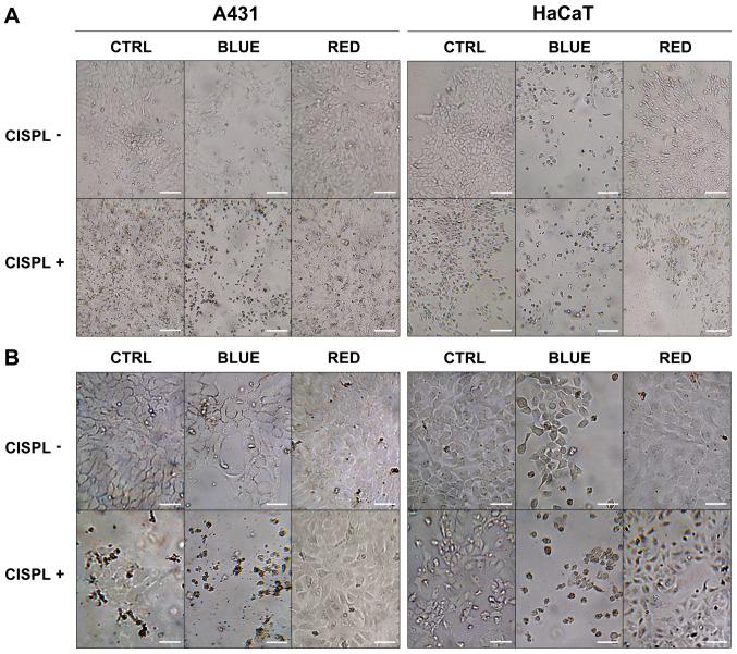
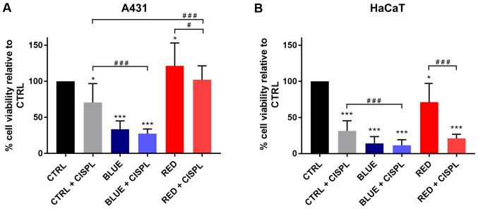
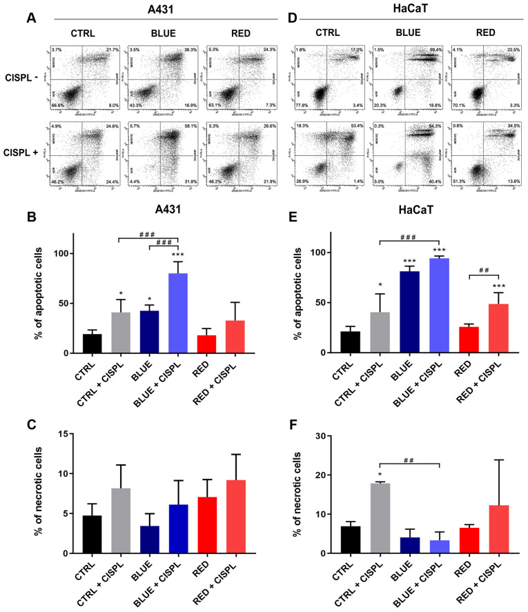
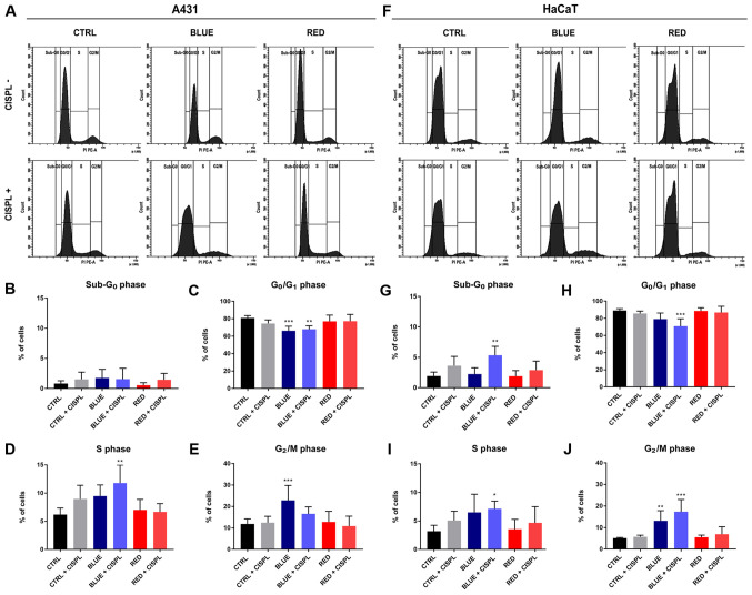
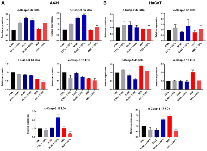
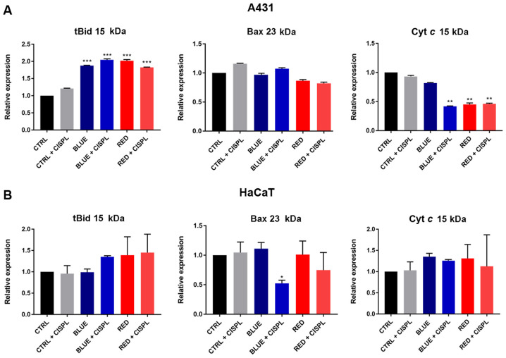
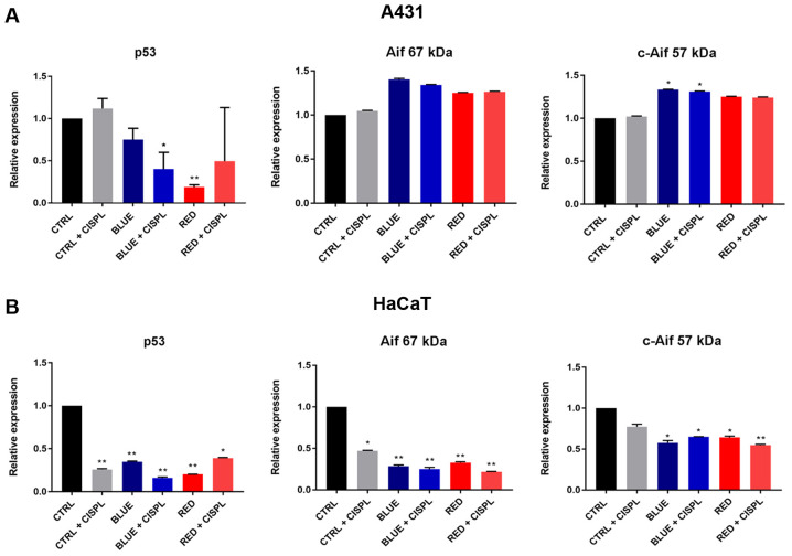
New approaches are being studied for the treatment of skin cancer. It has been reported that light combined with cisplatinum may be effective against skin cancer. In the present study, the effects of specific light radiations and cisplatinum on A431 cutaneous squamous cell carcinoma (cSCC) and HaCaT non‑tumorigenic cell lines were investigated. Both cell lines were exposed to blue and red light sources for 3 days prior to cisplatinum treatment. Viability, apoptosis, cell cycle progression and apoptotic‑related protein expression levels were investigated. The present results highlighted that combined treatment with blue light and cisplatinum was more effective in reducing cell viability compared with single treatments. Specifically, an increase in the apoptotic rate was observed when the cells were treated with blue light and cisplatinum, as compared to treatment with blue light or cisplatinum alone. Combined treatment with blue light and cisplatinum also caused cell cycle arrest at the S phase. Treatment with cisplatinum following light exposure induced the expression of apoptotic proteins in the A431 and HaCaT cell lines, which tended to follow different apoptotic mechanisms. On the whole, these data indicate that blue light combined with cisplatinum may be a promising treatment for cSCC.

Keywords: skin cancer; blue light; cisplatinum; A431 cells; HaCaT cells; LED light; light therapy; cSCC; apoptosis; cytotoxicity.


Conflict of interest statement

The authors declare that they have no competing interests.

Figures

Figure 1
Figure 1
Morphology of A431 and HaCaT cells exposed to LED, cisplatinum, and LED and cisplatinum combined. Representative images captured at magnifications of (A) x100 and (B) x200 magnification in A431 and HaCaT cell lines. Scale bars, (A) 60 μm and (B) 30 μm. LED, light-emitting diode.
Figure 2
Figure 2
Analysis of cell viability in (A) A431 and (B) HaCaT cell lines exposed to LED, cisplatinum, and LED and cisplatinum combined. Columns represent mean values and bars represent ± SD (n=3). *P<0.05 and ***P<0.001 vs. CTRL. #P<0.05 and ###P<0.001. LED, light-emitting diode; SD, standard deviation.
Figure 3
Figure 3
Apoptotic and necrotic effects of exposure of A431 and HaCaT cells to LED, cisplatinum, and LED and cisplatinum combined. Data indicate the percentage of apoptotic (Annexin V+) and necrotic (Annexin V-/PI+) cells of the (A-C) A431 and (D-F) HaCaT cell lines. Columns represent mean values and bars represent ± SD (n=3). *P<0.05, and ***P<0.001 vs. CTRL. ##P<0.01 and ###P<0.001. LED, light-emitting diode; SD, standard deviation.
Figure 4
Figure 4
Effect of exposure to LED, cisplatinum, and LED and cisplatinum combined on cell cycle progression in A431 and HaCaT cells. Data indicate the effect of every treatment on the different cell cycle phases of the (A-E) A431 and (F-J) HaCaT cell lines. Columns represent mean values and bars represent ± SD (n=3). *P<0.01, **P<0.001 and ***P<0.05 vs. CTRL. SD, standard deviation.
Figure 5
Figure 5
Analysis of apoptosis-related proteins Casp-9, Casp-8 and Casp-3 expression levels in cell lysates from (A) A431 and (B) HaCaT cells. Values were normalized to GAPDH. Columns represent mean values and bars represent ± SD (n=3). *P<0.05, **P<0.01 and ***P<0.001 vs. CTRL. Representative blot bands are presented in Fig. S1. SD, standard deviation; Casp, caspase.
Figure 6
Figure 6
Western blot analysis of apoptosis-related proteins Cyt c, Bax and tBid in (A) A431 and (B) HaCaT cells. Values were normalized to GAPDH. Columns represent mean values and bars represent ± SD (n=3). *P<0.05, **P<0.01 and ***P<0.001 vs. CTRL. Representative blot bands are presented in Fig. S1. tBid, truncated BH3 interacting domain death agonist; Bax, Bcl-2-associated X protein; Cyt c, cytochrome c; SD, standard deviation.
Figure 7
Figure 7
Western blot analysis of apoptosis-related proteins Aif and p53 in (A) A431 and (B) HaCaT cells. Values were normalized to GAPDH. Columns represent mean values and bars ± SD (n=3). *P<0.05 and **P<0.01 vs. CTRL. Representative blot bands are presented in Fig. S1. Aif, anti-apoptosis independent; SD, standard deviation.

Similar articles

Cited by

References

    1. Barolet D, Roberge CJ, Auger FA, Boucher A, Germain L. Regulation of skin collagen metabolism in vitro using a pulsed 660 nm LED light source: Clinical correlation with a single-blinded study. J Invest Dermatol. 2009;129:2751–2759. doi: 10.1038/jid.2009.186. - DOI - PubMed
    1. Mateus C. Cutaneous squamous cell carcinoma. Rev Prat. 2014;64:45–52. - PubMed
    1. Parekh V, Seykora JT. Cutaneous squamous cell carcinoma. Clin Lab Med. 2017;37:503–525. doi: 10.1016/j.cll.2017.06.003. - DOI - PubMed
    1. Smoller BR. Squamous cell carcinoma: From precursor lesions to high-risk variants. Mod Pathol. 2006;19(Suppl 2):S88–S92. doi: 10.1038/modpathol.3800509. - DOI - PubMed
    1. Palme CE, MacKay SG, Kalnins I, Morgan GJ, Veness MJ. The need for a better prognostic staging system in patients with metastatic cutaneous squamous cell carcinoma of the head and neck. Curr Opin Otolaryngol Head Neck Surg. 2007;15:103–106. doi: 10.1097/MOO.0b013e3280586723. - DOI - PubMed