

High neutralizing potency of swine glyco-humanized polyclonal antibodies against SARS-CoV-2
- PMID: 33576494
- PMCID: PMC8014652
- DOI: 10.1002/eji.202049072
High neutralizing potency of swine glyco-humanized polyclonal antibodies against SARS-CoV-2
Abstract
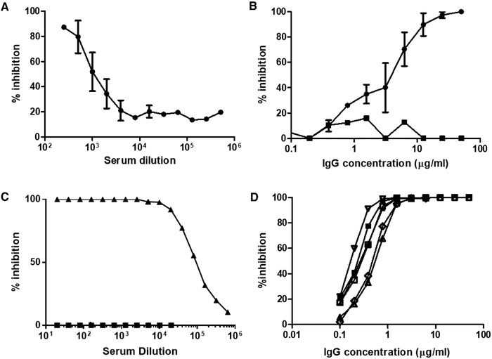
Heterologous polyclonal antibodies might represent an alternative to the use of convalescent plasma or monoclonal antibodies (mAbs) in coronavirus disease (COVID-19) by targeting multiple antigen epitopes. However, heterologous antibodies trigger human natural xenogeneic antibody responses particularly directed against animal-type carbohydrates, mainly the N-glycolyl form of the neuraminic acid (Neu5Gc) and the α1,3-galactose, potentially leading to serum sickness or allergy. Here, we immunized cytidine monophosphate-N-acetylneuraminic acid hydroxylase and α1,3-galactosyl-transferase (GGTA1) double KO pigs with the Severe acute respiratory syndrome coronavirus 2 (SARS-CoV-2) spike receptor binding domain to produce glyco-humanized polyclonal neutralizing antibodies lacking Neu5Gc and α1,3-galactose epitopes. Animals rapidly developed a hyperimmune response with anti-SARS-CoV-2 end-titers binding dilutions over one to a million and end-titers neutralizing dilutions of 1:10 000. The IgG fraction purified and formulated following clinical Good Manufacturing Practices, named XAV-19, neutralized spike/angiotensin converting enzyme-2 interaction at a concentration <1 μg/mL, and inhibited infection of human cells by SARS-CoV-2 in cytopathic assays. We also found that pig GH-pAb Fc domains fail to interact with human Fc receptors, thereby avoiding macrophage-dependent exacerbated inflammatory responses and a possible antibody-dependent enhancement. These data and the accumulating safety advantages of using GH-pAbs in humans warrant clinical assessment of XAV-19 against COVID-19.
Keywords: COVID-19; SARS-CoV-2; pig; polyclonal antibodies; spike.
© 2021 Wiley-VCH GmbH.
Conflict of interest statement
JR, PJR, CC, GE, EL, and BV are employees of Xenothera, a company developing glycol‐humanized polyclonal antibodies as those described in this manuscript, and OD, JPS, JMB, CG are cofounders of Xenothera. All other authors of this manuscript have no commercial or financial conflicts of interest.
Figures
Update of
-
High neutralizing potency of swine glyco-humanized polyclonal antibodies against SARS-CoV-2.bioRxiv [Preprint]. 2020 Oct 28:2020.07.25.217158. doi: 10.1101/2020.07.25.217158. bioRxiv. 2020. Update in: Eur J Immunol. 2021 Jun;51(6):1412-1422. doi: 10.1002/eji.202049072. PMID: 34013271 Free PMC article. Updated. Preprint.
Similar articles
-
High neutralizing potency of swine glyco-humanized polyclonal antibodies against SARS-CoV-2.bioRxiv [Preprint]. 2020 Oct 28:2020.07.25.217158. doi: 10.1101/2020.07.25.217158. bioRxiv. 2020. Update in: Eur J Immunol. 2021 Jun;51(6):1412-1422. doi: 10.1002/eji.202049072. PMID: 34013271 Free PMC article. Updated. Preprint.
-
XAV-19, a Swine Glyco-Humanized Polyclonal Antibody Against SARS-CoV-2 Spike Receptor-Binding Domain, Targets Multiple Epitopes and Broadly Neutralizes Variants.Front Immunol. 2021 Nov 15;12:761250. doi: 10.3389/fimmu.2021.761250. eCollection 2021. Front Immunol. 2021. PMID: 34868003 Free PMC article.
-
Evaluation of the safety and efficacy of XAV-19 in patients with COVID-19-induced moderate pneumonia: study protocol for a randomized, double-blinded, placebo-controlled phase 2 (2a and 2b) trial.Trials. 2021 Mar 9;22(1):199. doi: 10.1186/s13063-021-05132-9. Trials. 2021. PMID: 33750432 Free PMC article.
-
Structural Analysis of Neutralizing Epitopes of the SARS-CoV-2 Spike to Guide Therapy and Vaccine Design Strategies.Viruses. 2021 Jan 19;13(1):134. doi: 10.3390/v13010134. Viruses. 2021. PMID: 33477902 Free PMC article. Review.
-
Neutralizing antibody: a savior in the Covid-19 disease.Mol Biol Rep. 2022 Mar;49(3):2465-2474. doi: 10.1007/s11033-021-07020-6. Epub 2022 Jan 6. Mol Biol Rep. 2022. PMID: 34988889 Free PMC article. Review.
Cited by
-
Current Techniques of Gene Editing in Pigs for Xenotransplantation.Transpl Int. 2025 May 27;38:13807. doi: 10.3389/ti.2025.13807. eCollection 2025. Transpl Int. 2025. PMID: 40497031 Free PMC article. Review.
-
Tocilizumab administration for the treatment of hospitalized patients with COVID-19: A systematic review and meta-analysis.Respirology. 2021 Nov;26(11):1027-1040. doi: 10.1111/resp.14152. Epub 2021 Oct 3. Respirology. 2021. PMID: 34605114 Free PMC article.
-
The Road towards Polyclonal Anti-SARS-CoV-2 Immunoglobulins (Hyperimmune Serum) for Passive Immunization in COVID-19.Life (Basel). 2021 Feb 15;11(2):144. doi: 10.3390/life11020144. Life (Basel). 2021. PMID: 33671893 Free PMC article.
-
Therapeutic antibodies for COVID-19: is a new age of IgM, IgA and bispecific antibodies coming?MAbs. 2022 Jan-Dec;14(1):2031483. doi: 10.1080/19420862.2022.2031483. MAbs. 2022. PMID: 35220888 Free PMC article. Review.
-
XAV-19, un anti-variant de Sars-CoV-2, BQ.1.1 compris.Rev Francoph Lab. 2023 Mar;2023(550):6-7. doi: 10.1016/S1773-035X(23)00036-9. Epub 2023 Mar 2. Rev Francoph Lab. 2023. PMID: 36879983 Free PMC article. French. No abstract available.
References
-
- Casadevall, A. , Dadachova, E. and Pirofski, L. A. , Passive antibody therapy for infectious diseases. Nat. Rev. Microbiol. 2004. 2: 695–703. - PubMed
-
- Hsu, J. L. and Safdar, N. , Polyclonal immunoglobulins and hyperimmune globulins in prevention and management of infectious diseases. Infect. Dis. Clin. North Am. 2011. 25: 773–788. - PubMed
Publication types
MeSH terms
Substances
LinkOut - more resources
Full Text Sources
Other Literature Sources
Medical
Research Materials
Miscellaneous
