Review
. 2021 Apr;30(4):761-772.
doi: 10.1002/pro.4043. Epub 2021 Feb 24.

Structural and functional understanding of the toll-like receptors

Affiliations
Review

Structural and functional understanding of the toll-like receptors

Jinta Asami et al. Protein Sci. 2021 Apr.

Abstract

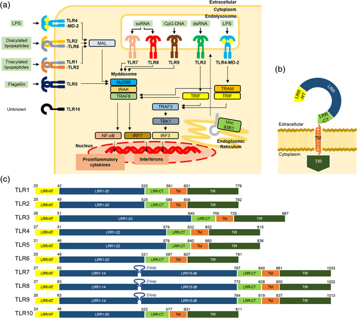
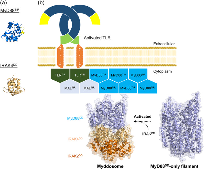
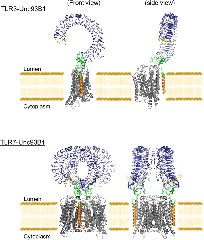
Recognition of invading pathogens by the innate immune system is essential to initiate antimicrobial responses and trigger adaptive immunity. This is largely mediated by an array of pattern-recognition receptor families that are essential for recognizing conserved molecular motifs characteristic of pathogenic microbes. One such family is the Toll-like receptors (TLRs). Activation of TLRs induces production of pro-inflammatory cytokines and type I interferons: the former triggers the synthesis of inflammatory mediators which cause fever, pain and other inflammation, and the latter mediates antiviral responses. Over the past decade, significant progress has been made in structural elucidation of TLRs in higher eukaryotes. The TLR structures with and without agonist and antagonist have been revealed by X-ray crystallography and cryo-electron microscopy studies, demonstrating the activated dimer formation induced by the agonistic ligand and the inhibition mechanism of the antagonistic ligand. Intracellular assembled structures and the TLR-chaperone complex are also reported. As the structural understanding of TLRs becomes better integrated with biochemical and immunological studies, a more comprehensive picture of their architectural and functional properties will emerge. This review summarizes recent advances in structural biological and mechanistic studies on TLRs.

Keywords: Myddosome; TIR; endolysosome; innate immunity; toll-like receptor.


Conflict of interest statement

The authors declare no competing financial interests.

Figures

FIGURE 1
FIGURE 1
TLR signaling and domain organization of TLRs. (a) Schematic illustration of TLR signaling pathway and downstream effector molecules. The ligand of each TLR is shown on the side, respectively. Nucleic‐acids sensing TLRs are trafficked from the endoplasmic reticulum to the endolysosome by Unc93B1. TLR signaling is initiated by the ligand recognition, followed by the interaction with adaptor proteins, such as MyD88, MAL, TRIF, and TRAM. Finally, the downstream signaling induces the expression of inflammatory cytokines or type I interferons. (b) Schematic representation of full‐length TLR. Extracellular domain is composed of the N‐terminal (LRR‐NT), LRRs and the C‐terminal (LRR‐CT) regions, followed by a single transmembrane segment (TM) and an intracellular TIR domain (TIR). (c) Schematic representation of full‐length human TLR1‐10. The color scheme is the same as that used in (b). LRRs are numbered. TLR7‐9 contain a characteristic Z‐loop between LRR14 and LRR15. Residues are numbered based on reported structures and amino‐acid sequence alignment information, 48 respectively. Residues of TLR10 are numbered based on UniProt
FIGURE 2
FIGURE 2
Agonist‐induced activated structures of TLRs. The representative activated dimer structures of TLR1‐TLR2‐Pam3CSK4 (PDB ID: 2Z7X), TLR2‐Diprovocim (PDB ID: 6NIG), TLR2‐TLR6‐Pam2CSK4 (PDB ID: 3A79), TLR3‐dsRNA (PDB ID: 3CIY), TLR4‐MD‐2‐LPS (PDB ID: 3FXI), TLR5‐FliC (PDB ID: 3 V47), TLR7‐guanosine‐polyU (PDB ID: 5GMF), TLR8‐ORN06 (PDB ID: 4R07), and TLR9‐DNA1668 (PDB ID: 3WPC). The color scheme is the same as that used in Figure 1b. The agonists are shown in orange. The gray regions in the TLR1‐TLR2‐Pam3CSK4, TLR2‐Diprovocim, TLR2‐TLR6‐Pam2CSK4, and TLR5‐FliC structures indicate the non‐TLR portions. All structural figures were generated using the PyMOL program 70
FIGURE 3
FIGURE 3
Antagonist‐bound inactivated structures of TLRs. (a) The structures of TLR4‐MD‐2‐Eritoran (PDB ID: 2Z65) and TLR9‐iDNA4084 (PDB ID: 3WPG). The antagonists are shown in orange. The gray region in the TLR4‐MD‐2‐Eritoran structure indicates the non‐TLR portion. (b) The structure of TLR8‐CU‐CPT8m (PDB ID: 5WYX) (left) and the density maps of TLR8‐antagonist (CU‐CPT8m) (PDB ID: 5WYX), unliganded TLR8 (PDB ID: 3W3G), and TLR8‐agonist (CL097) (PDB ID: 3W3J) (right). The antagonists in the structure are shown in orange. (c) The structure of TLR7‐Cpd‐7 (PDB ID: 6LW1) (left) and the density maps of TLR7‐antagonist (Cpd‐6) in open and closed forms (EMDB ID: EMD‐30000, EMD‐1000) (right). The antagonists are shown in orange. The color scheme of all the structures is the same as that used in Figure 1(b). Structural figures were generated using the PyMOL 70 or UCSF Chimera program 71
FIGURE 4
FIGURE 4
Intracellular assembled structures. (a) The structures of MyD88TIR (PDB ID: 4EO7) and IRAK4DD (PDB ID: 2A9I). BB loop of MyD88TIR is shown in yellow. (b) Model of TIR‐ and DD‐induced TLR signaling. The crystal structures of Myddosome (PDB ID: 3MOP) and MyD88DD‐only filament (PDB ID: 6I3N) are shown. MyD88DD‐only filaments can form Myddosome with IRAK4DD and IRAK2DD upon the ligand binding to TLRs. All structural figures were generated using the PyMOL program 70
FIGURE 5
FIGURE 5
Structures of TLR‐Unc93B1. The structures of TLR3‐Unc93B1 (PDB ID: 7C76) and TLR7‐Unc93B1 (PDB ID: 7CYN). The color scheme of TLRs is the same as that used in Figure 1b. Unc93B1 is shown in gray. All structural figures were generated using the PyMOL program 70

References

    1. Akira S, Uematsu S, Takeuchi O. Pathogen recognition and innate immunity. Cell. 2006;124:783–801. - PubMed
    1. Anderson KV, Bokla L, Nusslein‐Volhard C. Establishment of dorsal‐ventral polarity in the drosophila embryo: The induction of polarity by the toll gene product. Cell. 1985;42:791–798. - PubMed
    1. Lemaitre B, Nicolas E, Michaut L, Reichhart JM, Hoffmann JA. The dorsoventral regulatory gene cassette spatzle/toll/cactus controls the potent antifungal response in drosophila adults. Cell. 1996;86:973–983. - PubMed
    1. Michel T, Reichhart JM, Hoffmann JA, Royet J. Drosophila toll is activated by gram‐positive bacteria through a circulating peptidoglycan recognition protein. Nature. 2001;414:756–759. - PubMed
    1. Dinarello CA. Proinflammatory cytokines. Chest. 2000;118:503–508. - PubMed

Publication types

Substances

LinkOut - more resources