. 2021 Feb 1;62(2):13.
doi: 10.1167/iovs.62.2.13.

Molecular Epidemiology in 591 Italian Probands With Nonsyndromic Retinitis Pigmentosa and Usher Syndrome

Affiliations

Molecular Epidemiology in 591 Italian Probands With Nonsyndromic Retinitis Pigmentosa and Usher Syndrome

Leonardo Colombo et al. Invest Ophthalmol Vis Sci. .

Abstract

Purpose: To describe the molecular epidemiology of nonsyndromic retinitis pigmentosa (RP) and Usher syndrome (US) in Italian patients.

Methods: A total of 591 probands (315 with family history and 276 sporadics) were analyzed. For 155 of them, we performed a family segregation study, considering a total of 382 relatives. Probands were analyzed by a customized multigene panel approach. Sanger sequencing was used to validate all genetic variants and to perform family segregation studies. Copy number variants of selected genes were analyzed by multiplex ligation-dependent probe amplification. Four patients who tested negative to targeted next-generation sequencing analysis underwent clinical exome sequencing.

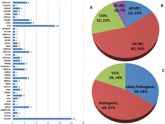
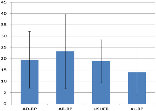
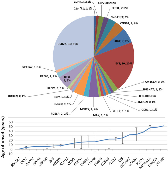
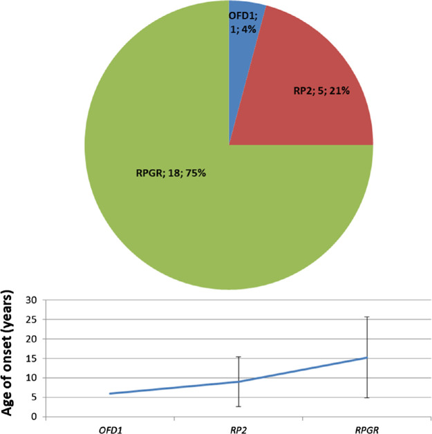
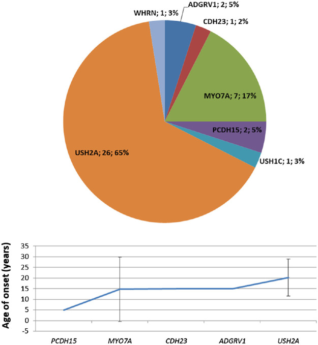
Results: The mean diagnostic yield of molecular testing among patients with a family history of retinal disorders was 55.2% while the diagnostic yield including sporadic cases was 37.4%. We found 468 potentially pathogenic variants, 147 of which were unpublished, in 308 probands and 66 relatives. Mean ages of onset of the different classes of RP were autosomal dominant RP, 19.3 ± 12.6 years; autosomal recessive RP, 23.2 ± 16.6 years; X-linked RP, 13.9 ± 9.9 years; and Usher syndrome, 18.9 ± 9.5 years. We reported potential new genotype-phenotype correlations in three probands, two revealed by TruSight One testing. All three probands showed isolated RP caused by biallelic variants in genes usually associated with syndromes such as PERCHING and Senior-Loken or with retinal dystrophy, iris coloboma, and comedogenic acne syndrome.

Conclusions: This is the largest molecular study of Italian patients with RP in the literature, thus reflecting the epidemiology of the disease in Italy with reasonable accuracy.


Conflict of interest statement

Disclosure: L. Colombo, None; P.E. Maltese, None; M. Castori, None; S. El Shamieh, None; C. Zeitz, None; I. Audo, None; A. Zulian, None; C. Marinelli, None; S. Benedetti, None; A. Costantini, None; S. Bressan, None; M. Percio, None; P. Ferri, None; A. Abeshi, None; M. Bertelli, None; L. Rossetti, None

Figures

Figure 1.
Figure 1.
Overview of the 147 unpublished variants: (A) distribution in genes, (B) disease distribution, and (C) classification of pathogenicity.
Figure 2.
Figure 2.
Prevalence of the four classes of RP based on genetic diagnosis in the whole population of 591 probands. (A) Probands, n = 591. (B) Relatives, n = 382.
Figure 3.
Figure 3.
Mean age of onset of the different classes of RP calculated on 214 genetically solved patients (probands and affected relatives). AD-RP, 19.5 ± 12.6 years; AR-RP, 23.2 ± 16.6 years; XL-RP, 13.9 ± 9.9 years; US, 18.9 ± 9.5 years. Bars indicate ± SD.
Figure 4.
Figure 4.
Genetic variants identified in AD-RP probands. Mean age of onset for each gene was calculated on 56 genetically solved patients (probands and affected relatives). Bars indicate ± SD.
Figure 5.
Figure 5.
Genetic variants identified in AR-RP probands. Mean age of onset for each gene was calculated on 93 genetically solved patients (probands and affected relatives). Bars indicate ± SD.
Figure 6.
Figure 6.
Genetic variants identified in XL-RP probands. Mean age of onset for each gene was calculated on 30 genetically solved patients (probands and affected relatives). Bars indicate ± SD.
Figure 7.
Figure 7.
Genetic variants identified in probands with US. Mean age of onset for each gene was calculated on 35 genetically solved patients (probands and affected relatives). Bars indicate ± SD.

References

    1. Berger W, Kloeckener-Gruissem B, Neidhardt J. The molecular basis of human retinal and vitreoretinal diseases. Prog Retin Eye Res. 2010; 29(5): 335–375. - PubMed
    1. Mrejen S, Audo I, Bonnel S, Sahel JA.. Retinitis pigmentosa and other dystrophies. Dev Ophthalmol. 2017; 58: 191–201. - PubMed
    1. Ziccardi L, Cordeddu V, Gaddini L, et al. .. Gene therapy in retinal dystrophies. Int J Mol Sci. 2019; 20(22): 5722. - PMC - PubMed
    1. Hamel C Retinitis pigmentosa. Orphanet J Rare Dis. 2006; 1: 40. - PMC - PubMed
    1. Colombo L, Montesano G, Sala B, et al. .. Comparison of 5-year progression of retinitis pigmentosa involving the posterior pole among siblings by means of SD-OCT: a retrospective study. BMC Ophthalmol. 2018; 18(1): 153. - PMC - PubMed

Publication types

MeSH terms

Substances