Review
. 2021 Apr;78(8):3969-3986.
doi: 10.1007/s00018-021-03762-5. Epub 2021 Feb 12.

Mitochondrial dynamics, positioning and function mediated by cytoskeletal interactions

Affiliations
Review

Mitochondrial dynamics, positioning and function mediated by cytoskeletal interactions

Mitali Shah et al. Cell Mol Life Sci. 2021 Apr.

Abstract

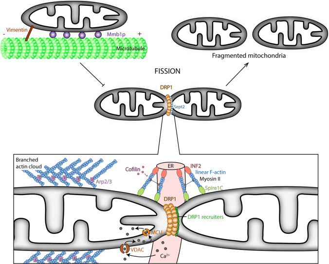
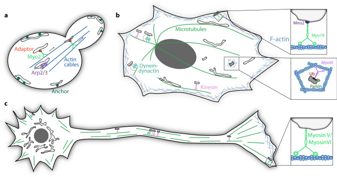
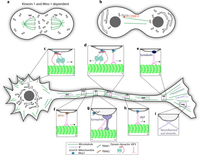
The ability of a mitochondrion to undergo fission and fusion, and to be transported and localized within a cell are central not just to proper functioning of mitochondria, but also to that of the cell. The cytoskeletal filaments, namely microtubules, F-actin and intermediate filaments, have emerged as prime movers in these dynamic mitochondrial shape and position transitions. In this review, we explore the complex relationship between the cytoskeleton and the mitochondrion, by delving into: (i) how the cytoskeleton helps shape mitochondria via fission and fusion events, (ii) how the cytoskeleton facilitates the translocation and anchoring of mitochondria with the activity of motor proteins, and (iii) how these changes in form and position of mitochondria translate into functioning of the cell.

Keywords: Cytoskeleton; Microtubules; Mitochondria; Mitochondrial dynamics; Molecular motors.


Conflict of interest statement

The authors declare that they have no conflict of interest with the contents of this article.

Figures

Fig. 1
Fig. 1
Overview of the players involved in effecting mitochondrial dynamics. Mitochondrial fission is mediated by DRP1, which is recruited by MFF, MID49 and MID51. Fission is inhibited when mitochondria are associated with microtubules. Mitochondrial fusion is brought about by L-OPA1, MFN1 and MFN2. F-actin and its associated proteins enable mitochondrial fission. S-OPA1 and MTP18 induce mitochondrial fission
Fig. 2
Fig. 2
The cytoskeleton and its associated proteins regulate mitochondrial dynamics. DRP1-mediated mitochondrial fission is inhibited when mitochondria are bound to microtubules via Mmb1p and vimentin. DRP1 binds directly to Sept2 and other recruitment factors at mitochondrial fission sites. F-actin aids mitochondrial fission at mitochondria-ER contact sites along with INF2, Spire1C and myosin II. Cofilin inhibits F-actin-mediated mitochondrial fission. DRP1-independent IMM constriction is enhanced through increased supply of calcium from the ER to the mitochondrial matrix via the VDAC and MCU. Arp2/3-mediated branched actin clouds also induce mitochondrial fission independent of the ER
Fig. 3
Fig. 3
F-actin and myosin motors participate in mitochondrial transport and anchoring. a The motor protein Myo2 and Arp2/3-mediated actin dynamics help transport mitochondria into the bud in S. cerevisiae. Mitochondria are additionally tethered along the cortex via anchor proteins. b In addition to the microtubule-based machinery, actin-based motors also participate in mitochondrial distribution. Myo19 associates with mitochondria either directly or via Miro proteins to organise mitochondria at the cell periphery and transport mitochondria on F-actin. Myosin VI is recruited onto mitophagy-destined mitochondria by ubiquitin, in order to isolate damaged mitochondria by triggering the formation of actin-cages around them. c Myosin V and VI have been implicated in activity-dependent anchorage of axonal mitochondria
Fig. 4
Fig. 4
Role of microtubules and intermediate filaments in the transport and anchorage of mitochondria. a Kinesin-1 transports mitochondria by the Miro-TRAK association on astral microtubules to the cytokinetic furrow during late anaphase. b Miro associates with CENP-F to transport mitochondria along growing microtubule plus-ends during late cytokinesis. c Dendritic mitochondria are predominantly transported on microtubules by the dynein–dynactin motor-adaptor complex which is recruited to mitochondria by the Miro1-TRAK2 association. d Axonal mitochondria are transported by both kinesin-1 and the dynein–dynactin complex, which are recruited to mitochondria predominantly by the Miro1-TRAK1 complex. e Syntabulin is an adaptor protein which recruits kinesin-1 to axonal mitochondria. f MFN1 recruits TRAK proteins to regulate mitochondrial transport on microtubules. g Syntaphilin anchors axonal mitochondria to microtubules in response to elevated Ca2+-levels by inhibiting kinesin-1 activity. h Increased glucose levels inhibit kinesin-1-dependent mitochondrial transport in neurons due to the activity of O-GlcNAc transferase on TRAK proteins. i Neurofilament and vimentin are the intermediate filaments involved in anchoring and organising mitochondria in neurons

References

    1. Kühlbrandt W. Structure and function of mitochondrial membrane protein complexes. BMC Biol. 2015;13:89. doi: 10.1186/s12915-015-0201-x. - DOI - PMC - PubMed
    1. Lee JH, Park A, Oh KJ, et al. The role of adipose tissue mitochondria: regulation of mitochondrial function for the treatment of metabolic diseases. Int J Mol Sci. 2019 doi: 10.3390/ijms20194924. - DOI - PMC - PubMed
    1. Duchen MR. Mitochondria and calcium: from cell signalling to cell death. J Physiol. 2000;529:57–68. doi: 10.1111/j.1469-7793.2000.00057.x. - DOI - PMC - PubMed
    1. Murphy MP. How mitochondria produce reactive oxygen species. Biochem J. 2009;417:1–13. doi: 10.1042/BJ20081386. - DOI - PMC - PubMed
    1. Wang C, Youle RJ. The role of mitochondria in apoptosis*. Annu Rev Genet. 2009;43:95–118. doi: 10.1146/annurev-genet-102108-134850. - DOI - PMC - PubMed

LinkOut - more resources