. 2021 Feb 10;7(2):129.
doi: 10.3390/jof7020129.

In Vitro and In Vivo Anti- Candida Activity and Structural Analysis of Killer Peptide (KP)-Derivatives

Affiliations

In Vitro and In Vivo Anti- Candida Activity and Structural Analysis of Killer Peptide (KP)-Derivatives

Tecla Ciociola et al. J Fungi (Basel). .

Abstract

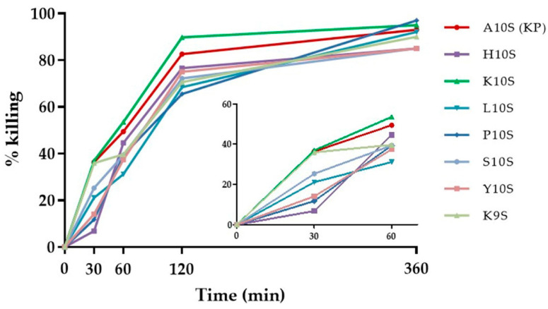
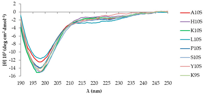
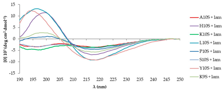
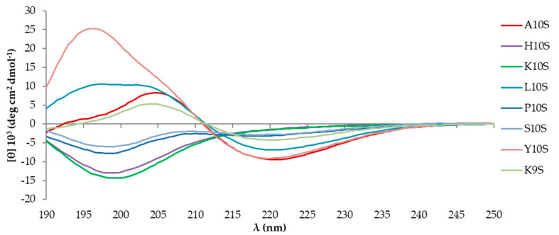
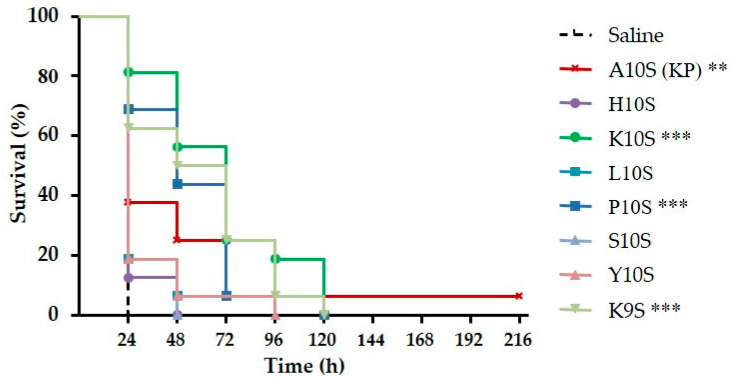
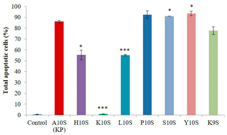
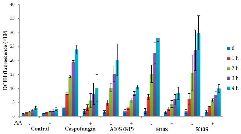
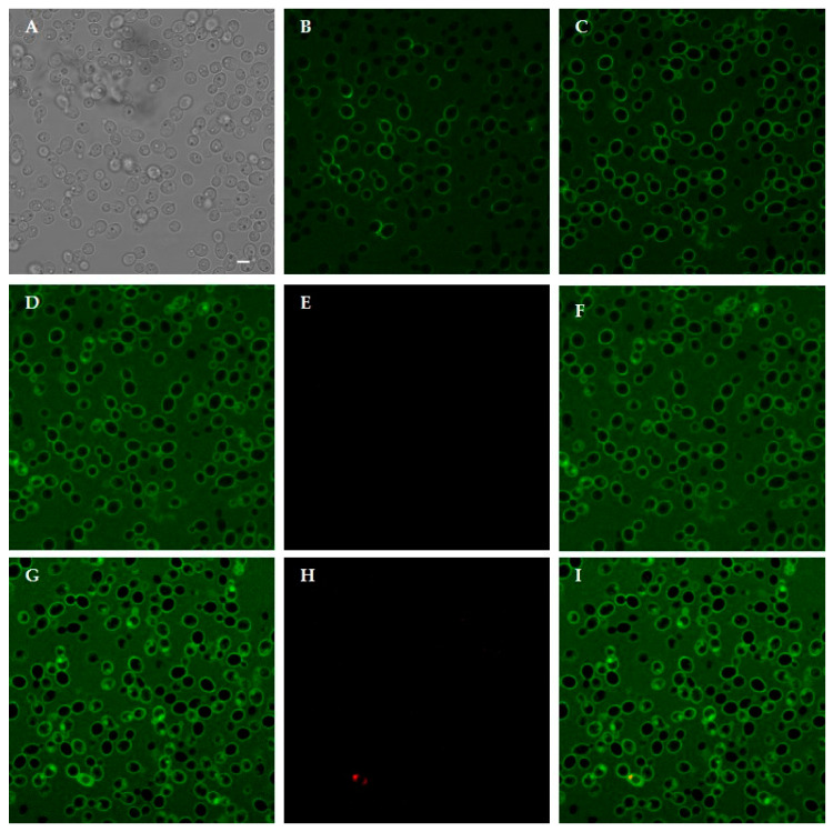
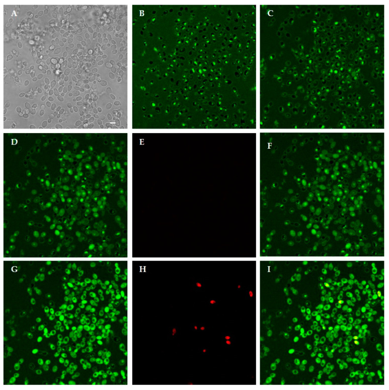
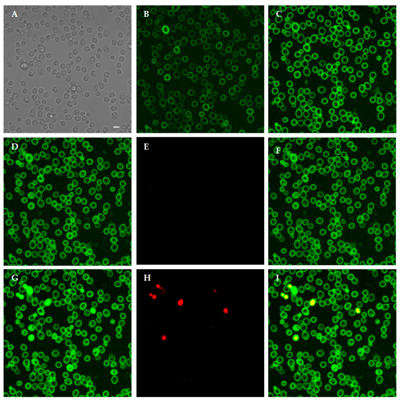
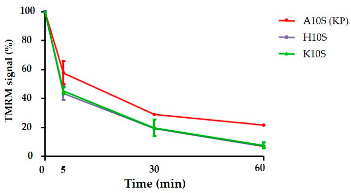
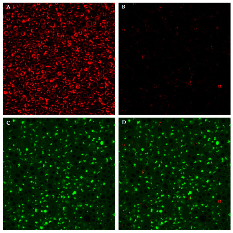
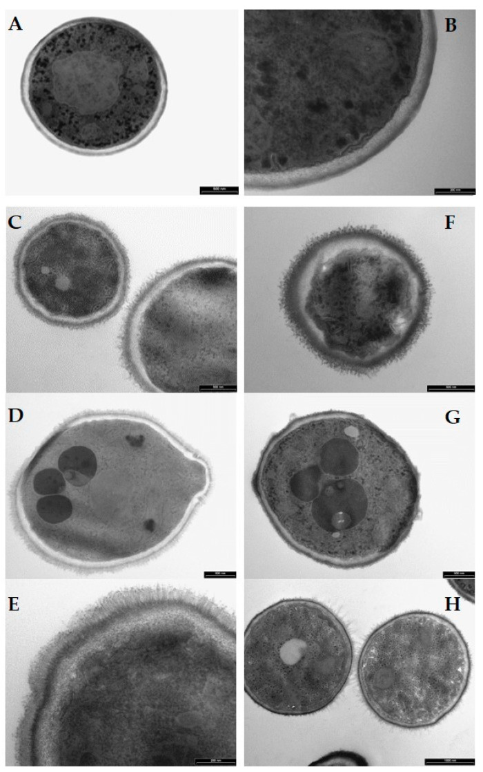
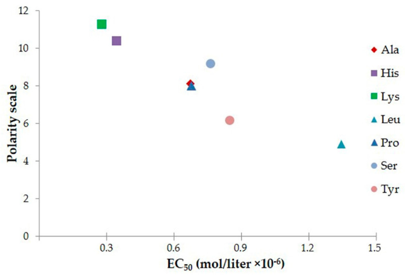
The previously described decapeptide AKVTMTCSAS (killer peptide, KP), derived from the variable region of a recombinant yeast killer toxin-like anti-idiotypic antibody, proved to exert a variety of antimicrobial, antiviral, and immunomodulatory activities. It also showed a peculiar self-assembly ability, likely responsible for the therapeutic effect in animal models of systemic and mucosal candidiasis. The present study analyzed the biological and structural properties of peptides derived from KP by substitution or deletion of the first residue, leaving unchanged the remaining amino acids. The investigated peptides proved to exert differential in vitro and/or in vivo anti-Candida activity without showing toxic effects on mammalian cells. The change of the first residue in KP amino acidic sequence affected the conformation of the resulting peptides in solution, as assessed by circular dichroism spectroscopy. KP-derivatives, except one, were able to induce apoptosis in yeast cells, like KP itself. ROS production and changes in mitochondrial transmembrane potential were also observed. Confocal and transmission electron microscopy studies allowed to establish that selected peptides could penetrate within C. albicans cells and cause gross morphological alterations. Overall, the physical and chemical properties of the first residue were found to be important for peptide conformation, candidacidal activity and possible mechanism of action. Small antimicrobial peptides could be exploited for the development of a new generation of antifungal drugs, given their relative low cost and ease of production as well as the possibility of devising novel delivery systems.

Keywords: Candida albicans; Galleria mellonella model; antifungal peptides; circular dichroism spectroscopy; confocal microscopy; electron microscopy; self-assembly peptides; structure-function relationship.


Conflict of interest statement

The authors declare no conflict of interest.

Figures

Figure 1
Figure 1
Time kinetics of C. albicans SC5314 killing. Peptides were used at their 2× EC50 value. The activity is expressed as percentage killing, calculated as: 100-(average number of CFUs in the peptide-treated group/average number of CFUs in the control group) × 100.
Figure 2
Figure 2
Far UV-CD spectra of 100 μM KP (A10S) and KP-derivatives acquired at 20 °C immediately after preparation of the starting aqueous solution (1 mM).
Figure 3
Figure 3
Far UV-CD spectra of 100 μM KP (A10S) and KP-derivatives acquired at 20 °C in the presence of laminarin (500 μM) immediately after preparation of the starting aqueous solution (1 mM).
Figure 4
Figure 4
Far UV-CD spectra of 100 μM KP (A10S) and KP-derivatives acquired at 20 °C 24 months after preparation of the starting aqueous solution (1 mM).
Figure 5
Figure 5
Therapeutic activity of KP-derivatives against experimental candidiasis in Galleria mellonella. Larvae were infected with 5 × 10⁵ cells of C. albicans SC5314 and treated with peptides (11 μmol/kg; single injection of 10 μL) or saline solution (control group). The survival curves of larvae treated with KP, K10S, P10S, and K9S were significantly different from that of the control group, as assessed by Mantel-Cox log rank test (** p < 0.01, *** p < 0.001).
Figure 6
Figure 6
Apoptotic effects of treatment with KP and its derivatives in Candida albicans SC5314 cells. Yeast cells were treated for 30 min with peptides at 2× their EC50 values. Data, expressed as percentage of apoptotic cells on the total gated cells, represent the mean ± standard deviation from at least two independent experiments (* p < 0.05, *** p < 0.001 vs. KP-treated cells).
Figure 7
Figure 7
Intracellular ROS production induced by treatment with KP and selected KP-derivatives. After incubation with (+) or without (−) ascorbic acid (AA), Candida albicans SC5314 cells were treated for 30 min with caspofungin (CAS, 20 µg/mL, positive control) and peptides KP, H10S, and K10S (20× EC50), then DCFH-DA was added and fluorescence intensity was monitored at different times. Data represent the mean ± standard deviation from at least two independent experiments.
Figure 8
Figure 8
Interaction between living Candida albicans cells and FITC-labeled KP. Confocal microscopy images of yeast cells incubated for 10 min and 150 min with the labeled peptide are shown in (B,C), respectively. KP bound to yeast cell wall. In (A), the same field is shown in light transmission. Peptide internalization was observed after 240 min: (D) FITC; (E) PI; (F) merge of (D,E) and increased over time leading to cell death, as demonstrated by PI internalization after 360 min: (G) FITC; (H) PI; (I) merge of (G,H). Bar, 5 μm.
Figure 9
Figure 9
Interaction between living Candida albicans cells and FITC-labeled H10S. Confocal microscopy images of yeast cells incubated for 10 min and 180 min with the labeled peptide are shown in (B,C), respectively. H10S was internalized in most yeast cells. In (A), the same field is shown in light transmission. Peptide internalization increased in viable cells after 240 min: (D) FITC; (E) PI; (F) merge of (D,E), eventually leading to cell death, as demonstrated by PI internalization at 360 min: (G) FITC; (H) PI; (I) merge of (G,H). Bar, 5 μm.
Figure 10
Figure 10
Interaction between living Candida albicans cells and FITC-labeled K10S. Confocal microscopy images of yeast cells incubated with the labeled peptide for 15 and 100 min are shown in (B,C), respectively. K10S was mainly bound to yeast cell wall. In (A), the same field is shown in light transmission. After 150 min, K10S entered viable yeast cells: (D) FITC; (E) PI; (F) merge of (D,E). Intensely fluorescent dead cells were observed after 180 min, as demonstrated by PI internalization: (G) FITC; (H) PI; (I) merge of (G,H). Bar, 5 μm.
Figure 11
Figure 11
Change in TMRM fluorescence intensity in Candida albicans SC5314 cells after peptide treatment. Data represent the mean ± standard deviation from three independent experiments.
Figure 12
Figure 12
Confocal images of living Candida albicans SC5314 cells pre-loaded with TMRM then treated for 30 min with FITC-labeled H10S: (B) TMRM; (C) FITC; (D) merge of (B,C), in comparison with untreated (control) cells: (A) TMRM. Bar, 5 µm.
Figure 13
Figure 13
Transmission electron microscopy images of Candida albicans cells treated for 60 min with selected KP-derivatives. (A,B): untreated (control) cells. (CE): H10S-treated cells. (FH): K10S-treated cells. Yeast cells treated with H10S and K10S (250 µM) revealed structural and morphological alterations. Bars = 500 nm (A,C,D,F,G), 1000 nm (H), 200 nm (B,E).
Figure 14
Figure 14
Correlation between polarity of the first residue and peptide EC50.

References

    1. Nii-Trebi N.I. Emerging and Neglected Infectious Diseases: Insights, Advances, and Challenges. Biomed. Res. Int. 2017;2017:5245021. doi: 10.1155/2017/5245021. - DOI - PMC - PubMed
    1. World Health Organization . Global Action Plan on Antimicrobial Resistance. WHO; Geneva, Switzerland: 2015. - PubMed
    1. Dodds D.R. Antibiotic resistance: A current epilogue. Biochem. Pharmacol. 2017;134:139–146. doi: 10.1016/j.bcp.2016.12.005. - DOI - PubMed
    1. Fisher M.C., Hawkins N.J., Sanglard D., Gurr S.J. Worldwide emergence of resistance to antifungal drugs challenges human health and food security. Science. 2018;360:739–742. doi: 10.1126/science.aap7999. - DOI - PubMed
    1. Brown E.D., Wright G.D. Antibacterial drug discovery in the resistance era. Nature. 2016;529:336–343. doi: 10.1038/nature17042. - DOI - PubMed

LinkOut - more resources