

Transfer Learning in Breast Cancer Diagnoses via Ultrasound Imaging
- PMID: 33578891
- PMCID: PMC7916666
- DOI: 10.3390/cancers13040738
Transfer Learning in Breast Cancer Diagnoses via Ultrasound Imaging
Abstract
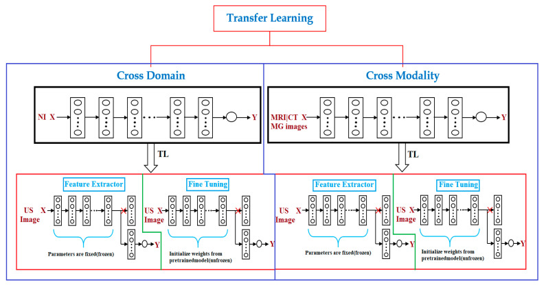
Transfer learning is a machine learning approach that reuses a learning method developed for a task as the starting point for a model on a target task. The goal of transfer learning is to improve performance of target learners by transferring the knowledge contained in other (but related) source domains. As a result, the need for large numbers of target-domain data is lowered for constructing target learners. Due to this immense property, transfer learning techniques are frequently used in ultrasound breast cancer image analyses. In this review, we focus on transfer learning methods applied on ultrasound breast image classification and detection from the perspective of transfer learning approaches, pre-processing, pre-training models, and convolutional neural network (CNN) models. Finally, comparison of different works is carried out, and challenges-as well as outlooks-are discussed.
Keywords: breast cancer; transfer learning; ultrasound.
Conflict of interest statement
The authors declare no conflict of interest.
Figures
References
-
- Smith N.B., Webb A. Ultrasound Imaging. In: Saltzman W.M., Chien S., editors. Introduction to Medical Imaging: Physics, Engineering and Clinical Applications. 6th ed. Volume 1. Cambridge University Press; Cambridge, UK: 2010. pp. 145–197. - DOI
-
- Gilbert F.J., Pinker-Domenig K. Diagnosis and Staging of Breast Cancer: When and How to Use Mammography, Tomosynthesis, Ultrasound, Contrast-Enhanced Mammography, and Magnetic Resonance Imaging. Dis. Chest Breast Heart Vessels. 2019;2019–2022:155–166. doi: 10.1007/978-3-030-11149-6_13. - DOI - PubMed
Publication types
Grants and funding
LinkOut - more resources
Full Text Sources
Other Literature Sources
Research Materials
