. 2021 Feb 10;13(4):526.
doi: 10.3390/polym13040526.

Curdlan-Chitosan Electrospun Fibers as Potential Scaffolds for Bone Regeneration

Affiliations

Curdlan-Chitosan Electrospun Fibers as Potential Scaffolds for Bone Regeneration

Clément Toullec et al. Polymers (Basel). .

Abstract

Polysaccharides have received a lot of attention in biomedical research for their high potential as scaffolds owing to their unique biological properties. Fibrillar scaffolds made of chitosan demonstrated high promise in tissue engineering, especially for skin. As far as bone regeneration is concerned, curdlan (1,3-β-glucan) is particularly interesting as it enhances bone growth by helping mesenchymal stem cell adhesion, by favoring their differentiation into osteoblasts and by limiting the osteoclastic activity. Therefore, we aim to combine both chitosan and curdlan polysaccharides in a new scaffold for bone regeneration. For that purpose, curdlan was electrospun as a blend with chitosan into a fibrillar scaffold. We show that this novel scaffold is biodegradable (8% at two weeks), exhibits a good swelling behavior (350%) and is non-cytotoxic in vitro. In addition, the benefit of incorporating curdlan in the scaffold was demonstrated in a scratch assay that evidences the ability of curdlan to express its immunomodulatory properties by enhancing cell migration. Thus, these innovative electrospun curdlan-chitosan scaffolds show great potential for bone tissue engineering.

Keywords: chitosan; curdlan; electrospinning; regenerative medicine; tissue engineering.


Conflict of interest statement

The authors declare no conflict of interest.

Figures
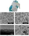
Figure 1
Figure 1
(a) Photography of the macroscopic aspect of the curdlan–chitosan scaffold as removed from the collector and SEM images of (b) electrospun chitosan membrane (Acceleration Voltage (AV) 20 keV), (c) electrospun curdlan–chitosan membrane (AV 20 keV), (d) cross section of electrospun curdlan–chitosan membrane (AV 3 keV), (e) electrospun curdlan–chitosan membrane after stabilization (AV 3 keV).
Figure 2
Figure 2
Size distribution of the fiber diameter for chitosan membrane in white and curdlan–chitosan membrane in black measured by image analysis with ImageJ on 100 randomly selected fibers.
Figure 3
Figure 3
ATR-FTIR spectra of curdlan and chitosan starting polymers and of the curdlan–chitosan fibers. (a) The full spectrum with peaks attribution [51]. (b) Zoom on the 1400 to 1700 cm−1 spectral range to identify characteristic peaks of both curdlan and chitosan of the blend spectrum (blue line denotes the 1530 cm−1 band specific to curdlan).
Figure 4
Figure 4
Swelling profile of the chitosan scaffolds (white dots) and curdlan–chitosan scaffolds (black dots) over time (* p < 0.05 et ** p < 0.01).
Figure 5
Figure 5
Degradation of the chitosan scaffolds (white) and curdlan–chitosan scaffolds (black) over time.
Figure 6
Figure 6
Metabolic activity over time of L929 mice fibroblast cells, control assays (dotted bars) and in the presence of pure chitosan scaffold (white bars) and curdlan–chitosan scaffold (black bars). Cell viability is expressed as % of the control evaluated at d0 (cells cultured in Dulbecco’s Modified Eagle’s Medium (DMEM) with 10% FBS). Values are presented as mean ± SEM.
Figure 7
Figure 7
Scratch assay with L929 mice fibroblast cells after 24 h in serum-free culture medium without any scaffold under microscope at 10× magnification, (a) with dissolved non-stabilized chitosan scaffold, (b) and with curdlan–chitosan scaffold (c). The images are representative of 3 independent experiments.
Figure 8
Figure 8
SEM images of L929 mice fibroblast cells on chitosan (a) and curdlan–chitosan (b) scaffold after 72 h of culture. The images are representative of 3 independent observations (AV 3 keV).

References

    1. Shegarfi H., Reikeras O. Bone transplantation and immune response. J. Orthop. Surg. 2009;2:206–211. doi: 10.1177/230949900901700218. - DOI - PubMed
    1. Yoshimoto H., Shin Y.M., Terai H., Vacanti J.P. A biodegradable nanofiber scaffold by electrospinning and its potential for bone tissue engineering. Biomaterials. 2003;24:2077–2082. doi: 10.1016/S0142-9612(02)00635-X. - DOI - PubMed
    1. Pape H.C., Evans A., Kobbe P. Autologous Bone Graft: Properties and Techniques. J. Orthop. Trauma. 2010;24:S36. doi: 10.1097/BOT.0b013e3181cec4a1. - DOI - PubMed
    1. Ratner B.D., Hoffman A.S., Schoen F.J., Lemons J.E. Biomaterials Science: An Introduction to Materials in Medicine. Elsevier; Amsterdam, The Netherlands: 2004.
    1. Donglu S. Introduction to Biomaterials. Tsinghua University Press; Beijing, China: 2005.

LinkOut - more resources