. 2021 Feb 10;11(2):260.
doi: 10.3390/biom11020260.

Unaltered Liver Regeneration in Post-Cholestatic Rats Treated with the FXR Agonist Obeticholic Acid

Affiliations

Unaltered Liver Regeneration in Post-Cholestatic Rats Treated with the FXR Agonist Obeticholic Acid

Lianne R de Haan et al. Biomolecules. .

Abstract

: In a previous study, obeticholic acid (OCA) increased liver growth before partial hepatectomy (PHx) in rats through the bile acid receptor farnesoid X-receptor (FXR). In that model, OCA was administered during obstructive cholestasis. However, patients normally undergo PHx several days after biliary drainage. The effects of OCA on liver regeneration were therefore studied in post-cholestatic Wistar rats. Rats underwent sham surgery or reversible bile duct ligation (rBDL), which was relieved after 7 days. PHx was performed one day after restoration of bile flow. Rats received 10 mg/kg OCA per day or were fed vehicle from restoration of bile flow until sacrifice 5 days after PHx. Liver regeneration was comparable between cholestatic and non-cholestatic livers in PHx-subjected rats, which paralleled liver regeneration a human validation cohort. OCA treatment induced ileal Fgf15 mRNA expression but did not enhance post-PHx hepatocyte proliferation through FXR/SHP signaling. OCA treatment neither increased mitosis rates nor recovery of liver weight after PHx but accelerated liver regrowth in rats that had not been subjected to rBDL. OCA did not increase biliary injury. Conclusively, OCA does not induce liver regeneration in post-cholestatic rats and does not exacerbate biliary damage that results from cholestasis. This study challenges the previously reported beneficial effects of OCA in liver regeneration in cholestatic rats.

Keywords: basolateral and canalicular transporters; bile acid metabolism; bile duct obstruction; bile salts; biliary obstruction; liver regeneration; partial hepatectomy; pharmacological intervention.


Conflict of interest statement

M.H. is the chief formulation officer at Nurish.Me LLC and Camelina Sun LTD and has equity in those companies (whose business activities are unrelated to the present work). There are no other conflict of interest.

Figures

Figure 1
Figure 1
Study design. rBDL, reversible bile duct ligation; Sham, sham surgery; OCA, obeticholic acid; Veh, vehicle; PHx, partial hepatectomy. n indicates the number of rats sacrificed at the indicated time point. † refers to animal sacrifice.
Figure 2
Figure 2
Obstructive cholestasis increases the total amount of serum and liver tissue bile acids (BAs) and bilirubin. Total amount of serum (A) and tissue BAs (B) and serum bilirubin levels (C) 1 day before PHx (t = −1 day), on the day of PHx (t = 0 days), and on 1, 3, and 5 days after PHx. Data points are given as mean ± SEM. Significance was assessed using a Mann–Whitney U test. * indicates p ≤ 0.05; ** indicates p ≤ 0.01; *** indicates p ≤ 0.001. Veh, vehicle; BDL, bile duct ligation; OCA, obeticholic acid.
Figure 3
Figure 3
Obstructive cholestasis does not induce proliferation prior to partial resection. (A) Dry liver weight and total (wet) liver weight 1 day before PHx (t = −1 day) expressed in g per 300 g body weight. (B) Number of mitotic cells per high-power field (HPF) in the area with the highest mitotic activity 1 day before PHx (t = −1 day). (C) mRNA expression levels in liver and ileum in the BDL group 6 days after BDL and 1 day before PHx (t = −1 day) expressed as fold-increase relative to the Sham group on t = −1 day. Significance was assessed using a Mann–Whitney U test. * indicates p < 0.05. Veh, vehicle; BDL, bile duct ligation.
Figure 4
Figure 4
(A) Number of mitotic cells per high-power field (HPF) in the area with the highest mitotic activity at the time of PHx (t = 0 days) and 1, 3, and 5 days after PHx. (B) Liver regrowth, expressed as percentage of dry remnant liver weight of the total dry liver weight, at the time of PHx (t = 0 days) and 1, 3, and 5 days after PHx. All data points are provided as mean ± SEM. Significance was assessed using a Mann–Whitney U test. An additional comparison was made between Sham-Veh vs. BDL-Veh. * indicates p < 0.05; ** indicates p < 0.01. BDL, bile duct ligation; Veh, vehicle; OCA, obeticholic acid.
Figure 5
Figure 5
mRNA expression of (A) liver and ileum genes involved in liver regeneration and (B) liver genes involved in BA homeostasis at the time of PHx (t = 0 days) and 1, 3, and 5 days after PHx. Transcript levels are expressed relative to the respective mean transcript levels in control rats on t = −1 day. Significance was assessed using a Kruskal–Wallis test with Dunn’s multiple comparisons test. * indicates p < 0.05; ** indicates p < 0.01; *** indicates p < 0.001. All data points represent mean ± SEM. Veh, vehicle; BDL, bile duct ligation; OCA, obeticholic acid.
Figure 6
Figure 6
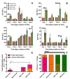
Obeticholic acid does not aggravate hepatobiliary injury caused by BDL. (AD) Alanine transaminase (ALT), aspartate transaminase (AST), alkaline phosphatase (ALP), and gamma-glutamyltransferase (gGT) serum levels expressed in units/liter. Data represent mean ± SEM. (E) Total inflammation score 1 day after PHx expressed as the sum of the scores for portal inflammation and lobular inflammation (Table S2). Data represent mean ± SD. (F) Types of fibrosis seen in each group 3 days after PHx. 0 = none; 1 = mild periportal fibrosis; 2 = moderate periportal fibrosis; 3 = bridging fibrosis (<50%). Significance was assessed using a Mann–Whitney U test (for t = −1 day) or a Kruskal–Wallis test with Dunn’s multiple comparisons test (for t = 0, 1, 3, and 5 days). * indicates p < 0.05; ** indicates p < 0.01; *** indicates p < 0.001. BDL, bile duct ligation; Veh, vehicle; OCA, obeticholic acid.
Figure 7
Figure 7
Serum fractional contribution of FXR agonists chenodeoxycholic acid and cholic acid, FXR antagonists tauromuricholic acids, and FXR-inert BAs to the total serum BA pool 1 day before PHx (t = −1 day), on the day of PHx (t = 0 days), and 1, 3, and 5 days after PHx. Significance in fractional contribution per BA between groups on the same time point was assessed using a Mann–Whitney U test (for t = −1 day) or a Kruskal–Wallis test with Dunn’s multiple comparisons test (for t = 0, 1, 3, and 5 days). * indicates p ≤ 0.05; ** indicates p ≤ 0.01. For intergroup analysis: *1, Sham-Veh vs BDL-Veh; *2, Sham-Veh vs BDL-OCA. BAs, bile acids; Veh, Vehicle; BDL, bile duct ligation; OCA, obeticholic acid.
Figure 8
Figure 8
Indocyanine green (ICG) plasma disappearance rate (PDR) expressed as percentage per minute. Significance was assessed using a using a Kruskal–Wallis test with Dunn’s multiple comparisons test. Data points represent mean ± SEM. Veh, vehicle; OCA, obeticholic acid; BDL, bile duct ligation.

References

    1. De Haan L., van der Lely S.J., Warps A.K., Hofsink Q., Olthof P.B., de Keijzer M.J., Lionarons D.A., Mendes-Dias L., Bruinsma B.G., Uygun K., et al. Post-hepatectomy liver regeneration in the context of bile acid homeostasis and the gutliver signaling axis. J. Clin. Transl. Res. 2018;4:1–46. doi: 10.18053/jctres.04.201801.001. - DOI - PMC - PubMed
    1. Michalopoulos G.K. Hepatostat: Liver regeneration and normal liver tissue maintenance. Hepatology. 2017;65:1384–1392. doi: 10.1002/hep.28988. - DOI - PubMed
    1. Taub R. Liver regeneration 4: Transcriptional control of liver regeneration. FASEB J. 1996;10:413–427. doi: 10.1096/fasebj.10.4.8647340. - DOI - PubMed
    1. Taub R. Liver regeneration: From myth to mechanism. Nat. Rev. Mol. Cell Biol. 2004;5:836–847. doi: 10.1038/nrm1489. - DOI - PubMed
    1. Hoekstra L.T., de Graaf W., Nibourg G.A.A., Heger M., Bennink R.J., Stieger B., van Gulik T.M. Physiological and Biochemical Basis of Clinical Liver Function Tests. Ann. Surg. 2013;257:27–36. doi: 10.1097/SLA.0b013e31825d5d47. - DOI - PubMed

Publication types

MeSH terms

LinkOut - more resources