Review
. 2021 Feb 12;22(1):11.
doi: 10.1186/s12860-021-00350-0.

Avian eggshell biomineralization: an update on its structure, mineralogy and protein tool kit

Affiliations
Review

Avian eggshell biomineralization: an update on its structure, mineralogy and protein tool kit

J Gautron et al. BMC Mol Cell Biol. .

Erratum in

Abstract

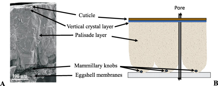
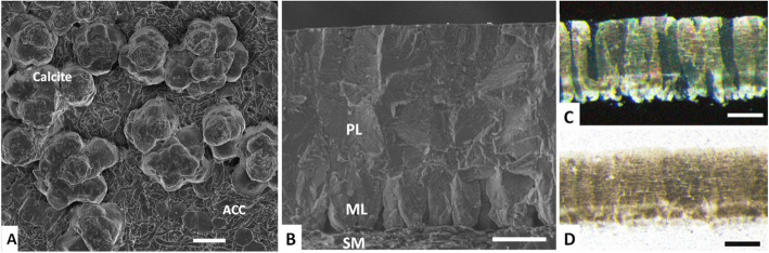
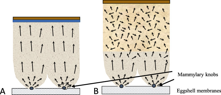
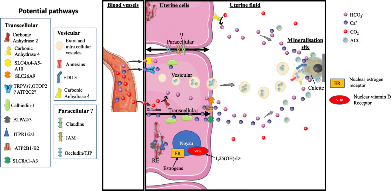
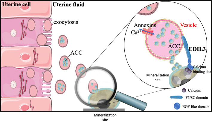
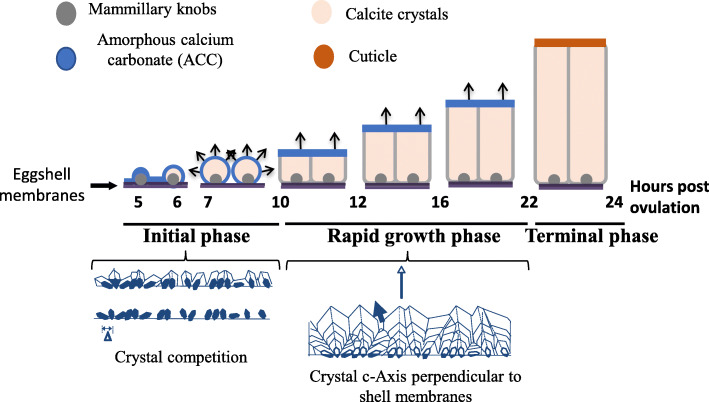
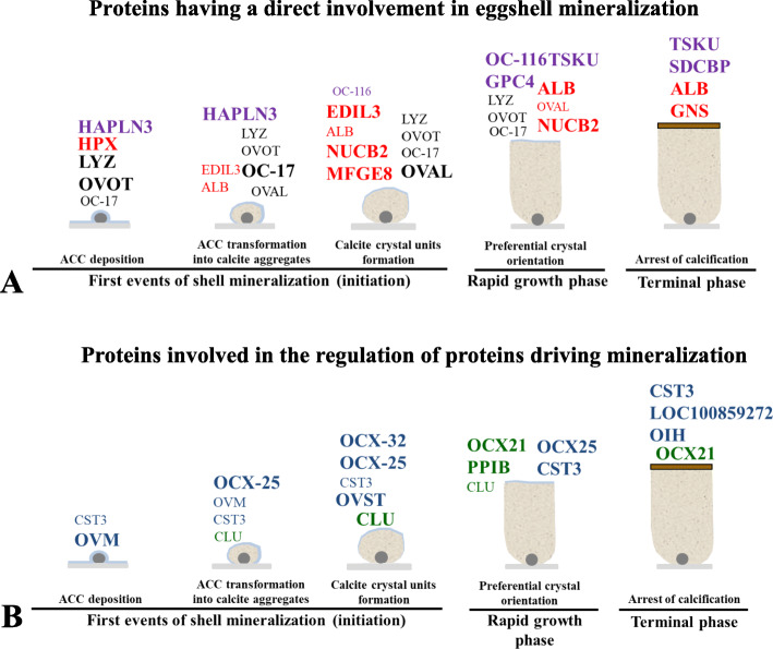
Background: The avian eggshell is a natural protective envelope that relies on the phenomenon of biomineralization for its formation. The shell is made of calcium carbonate in the form of calcite, which contains hundreds of proteins that interact with the mineral phase controlling its formation and structural organization, and thus determine the mechanical properties of the mature biomaterial. We describe its mineralogy, structure and the regulatory interactions that integrate the mineral and organic constituents during eggshell biomineralization. Main Body. We underline recent evidence for vesicular transfer of amorphous calcium carbonate (ACC), as a new pathway to ensure the active and continuous supply of the ions necessary for shell mineralization. Currently more than 900 proteins and thousands of upregulated transcripts have been identified during chicken eggshell formation. Bioinformatic predictions address their functionality during the biomineralization process. In addition, we describe matrix protein quantification to understand their role during the key spatially- and temporally- regulated events of shell mineralization. Finally, we propose an updated scheme with a global scenario encompassing the mechanisms of avian eggshell mineralization.

Conclusion: With this large dataset at hand, it should now be possible to determine specific motifs, domains or proteins and peptide sequences that perform a critical function during avian eggshell biomineralization. The integration of this insight with genomic data (non-synonymous single nucleotide polymorphisms) and precise phenotyping (shell biomechanical parameters) on pure selected lines will lead to consistently better-quality eggshell characteristics for improved food safety. This information will also address the question of how the evolutionary-optimized chicken eggshell matrix proteins affect and regulate calcium carbonate mineralization as a good example of biomimetic and bio-inspired material design.

Keywords: Amorphous calcium carbonate; Biomineralization; Calcite; Chicken; Eggshell; Extracellular vesicles; Ion supply; Matrix protein functions.


Conflict of interest statement

The authors declare not having competing interest.

Figures

Fig. 1
Fig. 1
Chicken eggshell structure. a Scanning electronic microphotograph cross-fractured eggshell. b Corresponding labeled drawing of the different layers of the eggshell
Fig. 2
Fig. 2
Eggshell ultrastructure and microstructure. a Scanning electron microscopy (SEM) image of an eggshell at an early stage of calcification (6 h post-ovulation.), showing aggregates of calcite crystals on mammillary knobs and ACC (amorphous calcium carbonate) flat-disk shaped particles on the shell membranes. b SEM image of cross-fractured eggshell showing the palisade layer (PL), mammillary layer (ML) and shell membranes (SM). Optical microscopy images of an eggshell cross-section: c as viewed under cross-polarized illumination, showing the columnar calcite crystal units of the mineral. d View under parallel light showing the distribution of the internal organic matter within the mineral. Scale bars are: a 10 μm; b 100 μm; c and d 200 μm
Fig. 3
Fig. 3
Comparison of schematic eggshell structure and crystal orientation in chicken (a) and in Guinea fowl (b) species. Black arrows represent the calcite crystal c-axes
Fig. 4
Fig. 4
Comprehensive model for calcium and carbonate transport to the uterine fluid during eggshell calcification. The three potential pathways for ion transfer through uterine cells are transcellular, vesicular and paracellular mechanisms. They could function asynchronously or in an integrated fashion. The major protein players in each pathway are indicated on the figure (left). Graphical elements were from Servier Medical Art (https://smart.servier.com/), licensed under a Creative Commons Attribution 3.0 Unported License. Adapted from [–25]
Fig. 5
Fig. 5
Proposed role for extracellular vesicles (EVs) in eggshell calcification. The EVs bud by exocytosis from the plasma membrane of the uterine cells. EVs transit the uterine fluid (UF) to deliver stabilized ACC (amorphous calcium carbonate) to the mineralization sites (MS). The passage of EV-encapsulated ACC avoids non-specific precipitation in the UF and provides stabilized ACC to the MS. Annexins allow calcium to penetrate into vesicles. EDIL3 (in bold) guides the EVs by targeting calcium to the mineralization front. Graphical elements were from Servier Medical Art (https://smart.servier.com/), licensed under a Creative Commons Attribution 3.0 Unported License. Adapted from Stapane [25]
Fig. 6
Fig. 6
Schematic representation of the different stages of eggshell deposition. Mineralization starts by massive accumulation of ACC at 5 h post-ovulation. ACC is transformed into ACC aggregates (6 h p.o.), and larger crystal units then form with their c-axes progressively perpendicular to the surface (7–10 h p.o.). During the growth phase (10–22 h p.o.), the compact layer (palisade layer) is deposited with all crystals oriented perpendicular to the surface. Two hours before oviposition, arrest of mineralization occurs and the thin organic cuticle layer is deposited. Arrows indicate the orientation of the c-axis of calcite crystals
Fig. 7
Fig. 7
Schematic representation of the sequential events of mineralization and major matrix proteins at five time points during shell mineralization. The font size is relative to relative protein levels in the eggshell extract. a Proteins having a direct involvement in shell mineralization. Black lettering for proteins with established role in biomineralization. Red lettering for proteins with calcium-binding domains. Purple for proteoglycans and proteoglycan-binding proteins. b Proteins involved in the regulation of proteins directing mineralization. Green lettering for chaperone proteins and blue lettering for proteases and protease inhibitors. Adapted from Marie and coworkers [30, 99]

References

    1. Hincke MT, Nys Y, Gautron J, Mann K, Rodriguez-Navarro AB, McKee MD. The eggshell: structure, composition and mineralization. Front Biosci. 2012;17:1266–1280. doi: 10.2741/3985. - DOI - PubMed
    1. Nys Y, Hincke MT, Arias JL, Garcia-Ruiz JM, Solomon SE. Avian eggshell mineralization. Poult Avian Biol Rev. 1999;10(3):143–166.
    1. Sauveur B, Derevier M. Reproduction des volailles et production d'oeufs Quae edn. Paris: INRA; 1988.
    1. Solomon SE. Egg and egg quality. London, England: Wolfe publishing; 1991.
    1. Panheleux M, Bain M, Fernandez MS, Morales I, Gautron J, Arias JL, Solomon SE, Hincke M, Nys Y. Organic matrix composition and ultrastructure of eggshell: a comparative study. Brit Poultry Sci. 1999;40(2):240–252. doi: 10.1080/00071669987665. - DOI - PubMed