Case Reports
. 2021 Feb 13;15(1):58.
doi: 10.1186/s13256-021-02676-x.

Anomalous plantar intrinsic foot muscle attaching to the medial longitudinal arch: possible mechanism for medial nerve entrapment: a case report

Affiliations
Case Reports

Anomalous plantar intrinsic foot muscle attaching to the medial longitudinal arch: possible mechanism for medial nerve entrapment: a case report

R Claire Aland et al. J Med Case Rep. .

Abstract

Background: Muscular variations are potentially symptomatic and may complicate imaging interpretation. Intrinsic foot musculature and extrinsic tendon insertion variations are common. Distinct supernumerary muscles are rare. We report a novel anomalous intrinsic foot muscle on the medial longitudinal arch.

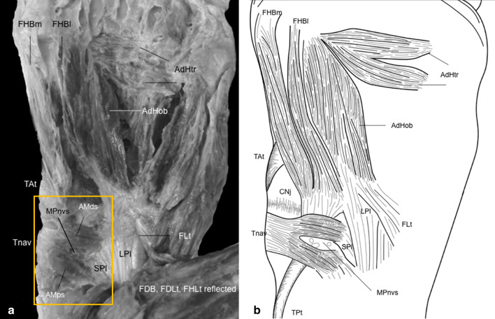
Case presentation: An accessory muscle was encountered on the medial arch of the right foot of a 78-year-old white male cadaver, between layers two and three of the foot intrinsics. It did not appear to be a slip or variant of a known foot muscle. This muscle consisted of two slips that ran transversely on the plantar aspect of the medial arch, crossing the medial transverse tarsal joint and attaching to the tuberosity of the navicular, the short and long plantar ligaments, and spring ligament.

Conclusions: The medial plantar vessels and nerve passed from deep to superficial between the two slips, and this suggests a possible location for medial nerve entrapment.

Keywords: Anatomical variation; Case report; Medial longitudinal arch; Medial nerve entrapment; Plantar intrinsic muscle.


Conflict of interest statement

The authors declare that they have no competing interests.

Figures

Fig. 1
Fig. 1
Anomalous muscle slips attach to the medial longitudinal arch: a photograph; b schematic drawing. Abductor hallucis (AbH) is removed, and the flexor digitorum longus (FDL) and flexor hallucis longus (FHL) tendons and the flexor digitorum brevis (FDB) have been reflected. AdHob Adductor hallucis oblique head; AdHtr adductor hallucis transverse head; AMds anomalous muscle distal slip; Amps anomalous muscle proximal slip; CNj cuneonavicular joint; FDB flexor digitorum brevis FDLt flexor digitorum longus tendon; FHLt reflected flexor hallucis longus tendon, reflected; FLt fibularis longus tendon; LPl long plantar ligament; MPnvs medial plantar nerves and vessels, SPl short plantar ligament, Tat tibialis anterior tendon, Tnav navicular tuberosity, TPt tibialis posterior tendon

References

    1. Agawany AE, Meguid EA. Mode of insertion of the abductor hallucis muscle in human feet and its arterial supply. Folia Morphol. 2010;69:54–61. - PubMed
    1. Arakawa T, Tokita K, Miki A, Terashima T. Anatomical study of human adductor hallucis muscle with respect to its origin and insertion. Ann Anat. 2003;185(6):585–592. doi: 10.1016/S0940-9602(03)80133-4. - DOI - PubMed
    1. Boyer EL. The musculature of the inferior extremity of the orang-utan, Simia satyrus. Am J Anat. 1935;56:192–256. doi: 10.1002/aja.1000560203. - DOI
    1. Brenner E. Insertion of the abductor hallucis muscle in feet with and without hallux valgus. Anat Rec. 1999;254:429–434. doi: 10.1002/(SICI)1097-0185(19990301)254:3<429::AID-AR14>3.0.CO;2-5. - DOI - PubMed
    1. Brooks HSJ. On the short muscles of the pollex and hallux of the anthropoid apes, with special reference to the opponens hallucis. J Anat Physiol. 1887;22:78–95. - PMC - PubMed

Publication types