Review
. 2021:149:321-387.
doi: 10.1016/bs.acr.2020.10.005. Epub 2020 Dec 9.

Intrahepatic cholangiocarcinoma: Morpho-molecular pathology, tumor reactive microenvironment, and malignant progression

Affiliations
Review

Intrahepatic cholangiocarcinoma: Morpho-molecular pathology, tumor reactive microenvironment, and malignant progression

Alphonse E Sirica et al. Adv Cancer Res. 2021.

Abstract

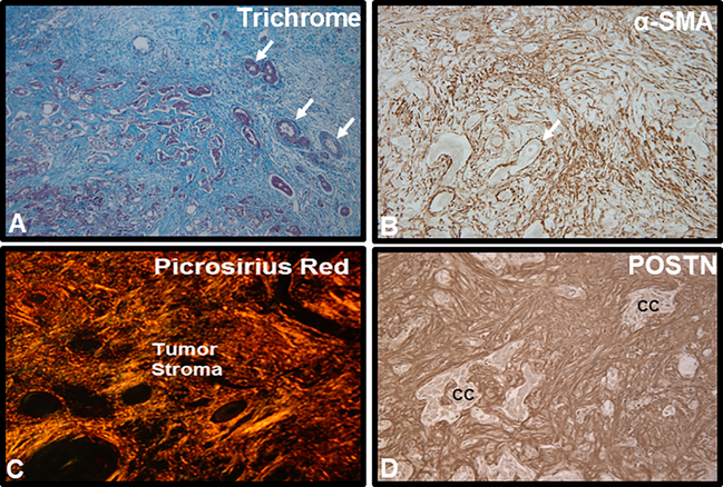
Intrahepatic cholangiocarcinoma (iCCA) is a relatively rare, but highly lethal and biologically complex primary biliary epithelial cancer arising within liver. After hepatocellular carcinoma, iCCA is the second most common primary liver cancer, accounting for approximately 10-20% of all primary hepatic malignancies. Over the last 10-20 years, iCCA has become the focus of increasing concern largely due to its rising incidence and high mortality rates in various parts of the world, including the United States. The challenges posed by iCCA are daunting and despite recent progress in the standard of care and management options for iCCA, the prognosis for this cancer continues to be dismal. In an effort to provide a framework for advancing our understanding of iCCA malignant aggressiveness and therapy resistance, this review will highlight key etiological, biological, molecular, and microenvironmental factors hindering more effective management of this hepatobiliary cancer. Particular focus will be on critically reviewing the cell origins and morpho-molecular heterogeneity of iCCAs, providing mechanistic insights into high risk fibroinflammatory cholangiopathies associated with iCCA development, and notably discussing the deleterious role played by the tumor reactive desmoplastic stroma in regulating iCCA malignant progression, lymphangiogenesis, and tumor immunobiology.

Keywords: Cancer-associated fibroblasts; Extracellular matrix; Fibroinflammatory risk conditions; Immune milieu; M2 macrophages; Morpho-molecular classification; Periostin; Therapeutic targeting; Transforming growth factor-β; Tumor reactive microenvironment.


Conflict of interest statement

Conflict of interest statement The authors (A.E.S., M.S., and M.C.) have no financial or personal disclosures relevant to this manuscript.

Figures

Figure 1.
Figure 1.
Representative histological images illustrating characteristic features of the desmoplastic microenvironment in human iCCA. A. Masson trichrome staining of a moderately to poorly-differentiated mass-forming iCCA demonstrating the tumor to be largely comprised of a prominent desmoplastic stroma strongly stained for collagen (blue staining). Arrows point to representative small clusters of cholangiocarcinoma. B. CAFs comprising the vast majority of cell types populating the desmoplastic stroma of iCCA are seen to be strongly immunoreactive for α- smooth muscle actin (α- SMA), a biomarker of myofibroblast differentiation, whereas cholangiocarcinoma cells (arrow) are negatively stained for α- SMA. C. Picrosirius red staining for collagen (orange-staining under polarized light) typically reveals the extracellular matrix of desmoplastic CCA to be comprised of thick collagen fiber bundles that are largely comprised of collagen type I. D. Immunostaining for matricellular periostin (Postn), produced by α-SMA+CAFs and which has a binding site for collagen, is exclusively localized to the desmoplastic stroma of iCCA. cc, cholangiocarcinoma. Increased numbers of α-SMA+CAFs within the iCCA microenvironment together with corresponding strong stromal immunoreactivity for Postn have been shown to be predictors of poor survival outcomes for iCCA patients following curative-intent surgical resection.
Figure 2.
Figure 2.
Relationship between mesothelin (Msln) immunostaining pattern, disruption of polarized morphogenesis, and invasiveness in a syngeneic orthotopic rat tumor model and corresponding 3-dimensional (3-D) organotypic culture model of cholangiocarcinoma progression. A. Immunohistochemical demonstration of luminal Msln immunoreactivity expressed in differentiated cholangiocarcinoma ductal structures (white arrows) contrasted with diffuse cell membrane/cytoplasmic Msln immunoreactivity (black arrows) expressed in invasive cholangiocarcinoma emanating from the ductal structures in an orthotopic rat iCCA (TDECC iCCA). B. Phase contrast image of viable spheroids and well-differentiated ductal-like structures with distinct lumens (L) formed in 3-D organotypic culture of a cholangiocarcinoma cell strain (TDECC cells) derived from rat TDECC iCCA. C. Hematoxylin & eosin stained section demonstrating polarized ductal-like structure formed from rat TDECC cholangiocarcinoma cells in 3-D culture. D. In the absence of CAFs, polarized ductal-like structures formed from TDECC cholangiocarcinoma cells in 3-D culture exhibit strong luminal surface immunoreactivity for Msln (white arrow heads). E. In sharp contrast, polarized morphogenesis is dramatically disrupted when TDECC cholangiocarcinoma cells are maintained in 3-D co-cultured with α- SMA+CAFs, also derived from a rat TDECC iCCA. Under these co-culture conditions, the cholangiocarcinoma cells can organized into hyperproliferative, invasive structures exhibiting diffuse cell membrane/cytoplasmic immunoreactivity for Msln (black arrows). In both the orthotopic tumor and organotypic culture models, a more heavily glycosylated 50-kDa form of Msln was demonstrated to be associated with the apical surface pattern of expression of Msln, whereas a 40-kDA Msln form was shown to be associated with the diffuse cell membrane/cytoplasmic immunostaining pattern. Data based on results presented in Manzanares et al., 2018. Images in 2A and 2E are slightly modified versions of those published as Figure 2B and Figure 7B, respectively, in Manzanares et al, 2018.

References

    1. Abou-Alfa GK, Macarulla T, Javle MM, Kelley RK, Lubner SJ, Adeva J, et al. (2020a). Ivosidenib in IDH1-mutant, chemotherapy-refractory cholangiocarcinoma (ClarlDHy): a multicentre, randomized, double-blind, placebo-controlled, phase 3 study. Lancet Oncology, 21: 796–807. - PMC - PubMed
    1. Abou-Alfa GK, Sahai V, Hollebecque A, Vaccaro G, Melisi D, Al-Rajabi R, et al. (2020b). Pemigatinib for previously treated, locally advanced or metastatic cholangiocarcinoma: a multicentre, open-label, phase 2 study. Lancet Oncology, 21: 671–684. - PMC - PubMed
    1. Affo S, Yu L-Y, & Schwabe RF (2017). The role of cancer-associated fibroblasts and fibrosis in liver cancer. Annual Review of Pathology: Mechanisms of Disease, 12: 153–186. - PMC - PubMed
    1. Ahrendt SA, Rashid A, Chow JT, Eisenberger CF, Pitt HA, & Sidransky D (2000). p53 overexpression and K-ras gene mutations in primary sclerosing cholangitis-associated biliary tract cancer. Journal of Hepato-Biliary-Pancreatic Surgery, 7: 426–431. - PubMed
    1. Aishima S, Taguchi K, Sugimachi K, Asayama Y, Nishi H, Shimada M, et al. (2002). The role of thymidine phosphorylase and thrombospondin-1 in angiogenesis and progression of intrahepatic cholangiocarcinoma. International Journal of Surgical Pathology, 10: 47–56. - PubMed

Publication types