. 2021 Feb 12;7(7):eabf1798.
doi: 10.1126/sciadv.abf1798. Print 2021 Feb.

Anti-USAG-1 therapy for tooth regeneration through enhanced BMP signaling

Affiliations

Anti-USAG-1 therapy for tooth regeneration through enhanced BMP signaling

A Murashima-Suginami et al. Sci Adv. .

Abstract

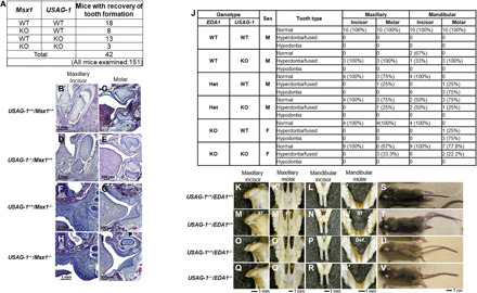
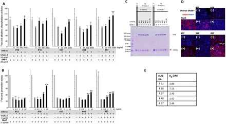
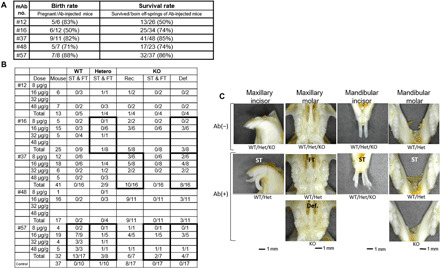
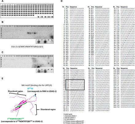
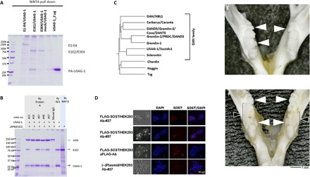
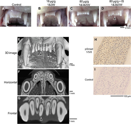
Uterine sensitization-associated gene-1 (USAG-1) deficiency leads to enhanced bone morphogenetic protein (BMP) signaling, leading to supernumerary teeth formation. Furthermore, antibodies interfering with binding of USAG-1 to BMP, but not lipoprotein receptor-related protein 5/6 (LRP5/6), accelerate tooth development. Since USAG-1 inhibits Wnt and BMP signals, the essential factors for tooth development, via direct binding to BMP and Wnt coreceptor LRP5/6, we hypothesized that USAG-1 plays key regulatory roles in suppressing tooth development. However, the involvement of USAG-1 in various types of congenital tooth agenesis remains unknown. Here, we show that blocking USAG-1 function through USAG-1 knockout or anti-USAG-1 antibody administration relieves congenital tooth agenesis caused by various genetic abnormalities in mice. Our results demonstrate that USAG-1 controls the number of teeth by inhibiting development of potential tooth germs in wild-type or mutant mice missing teeth. Anti-USAG-1 antibody administration is, therefore, a promising approach for tooth regeneration therapy.


Figures

Fig. 1
Fig. 1. Recovery of tooth formation in double KO mice with congenital tooth agenesis and supernumerary teeth.
(A) Number of mice with indicated genotypes. (B to I) Frontal hematoxylin and eosin–stained sections of the left maxillary incisor and third molar (M3) in USAG-1−/−/Msx1−/− mice immediately after birth. (J) Summary of tooth phenotypes in 8-month-old F2 generation EDA1/USAG-1 double-mutant mice. (K to R′) Representative tooth phenotypes in dry skulls of 8-month-old F2 generation EDA1/USAG-1 double-mutant mice. (S to V) Ear hair, tail hair, and tail tip phenotypes. ST, supernumerary teeth; FT, fused teeth; Def., defect of teeth. Photo credit: H. Kiso, Kyoto University.
Fig. 2
Fig. 2. In vitro analyses of five types of USAG-1–neutralizing antibodies (#12, #16, #37, #48, and #57).
(A) Neutralization of the antagonistic activity of BMP signaling by USAG-1 antibodies as assessed by alkaline phosphatase assay. (B) Neutralization of the antagonistic activity of Wnt signaling by USAG-1 antibodies in a Wnt reporter assay. (C) Binding between anti–USAG-1 antibody and human/mouse-PA-USAG-1 protein in pull-down assays. (D) Immunocytochemistry of human embryonic kidney (HEK) 293–expressing FLAG-tagged human USAG-1 protein. (E) KD values of each USAG-1 antibody toward the mouse USAG-1 protein. mAb, monoclonal antibody; Ab, antibody; DAPI, 4′,6-diamidino-2-phenylindole.
Fig. 3
Fig. 3. Recovery of tooth defects in EDA1 mutant mice and whole tooth regeneration upon administration of USAG-1–neutralizing antibodies.
(A) Offspring birth and survival rates. (B) Summary of incidence of tooth phenotypes, including supernumerary teeth and fused teeth (ST and FT), recovery of teeth (Rec.), and defect of teeth (Def.). (C) Representative tooth phenotype in dry skulls of 8-month-old mice. Photo credit: A. Murashima-Suginami, Kyoto University.
Fig. 4
Fig. 4. Epitope mapping of neutralizing USAG-1 antibodies #37 and #57.
(A) The pattern of 14-mer peptide spots on the membrane from A1 to F19 [recombinant USAG-1 protein from: 1, mammalian cells; 2, Baculovirus; and 3, E. coli; USAG-1 antibodies (A) #37 and (B) #57]. (B) The peptide array probed with USAG-1 antibody #37. (C) The peptide array probed with USAG-1 antibody #57. (D) The number and sequence of the 14-mer peptide from A1 to F19. (E) Suggested 3D nuclear magnetic resonance structure model of mouse USAG-1 protein. Green, the epitope for USAG-1 antibody #37. Sky blue, the binding site for LRP5/6 (NX1 motif).
Fig. 5
Fig. 5. USAG-1–neutralizing antibodies sufficient for generating a whole tooth (#37 and #57) inhibit the antagonistic function of BMP but not Wnt signaling.
(A) Interaction between the extracellular E1/E2 domain of LRP6 and mouse USAG-1 protein. (B) Blocking of the interaction between the extracellular domain of LRP6 E1/E2 and mouse USAG-1 protein by USAG-1 antibodies (#16). IgG, immunoglobulin G. (C) Dendrogram of DAN family proteins that are BMP antagonists. (D) Cross-reactivity of the USAG-1 antibody #57 to the SOST protein expressed in HEK293 cells. (E) Phenotype of the mandibular molar in the dry skull of a USAG-1−/− mouse. (F) Phenotype of the mandibular molar in the dry skull of a USAG-1−/− mouse administered a mix of USAG-1 antibodies (#12, #16, #37, #48, and #57). White arrowheads indicate supernumerary teeth; black arrowheads indicate enlarged fused teeth. Photo credit: A. Murashima-Suginami, Kyoto University.
Fig. 6
Fig. 6. Supernumerary tooth of maxillary incisors of ferrets upon administration of USAG-1–neutralizing antibody #37.
(A to D) Maxillary incisors of ferrets to different doses of administration USAG-1–neutralizing antibody #37. (E to G) Micro-computed tomography (micro-CT) image of Fig. 6D. (H and I) Immunolocalization of phosphorylated Smad1/5/8 (pSmad1/5/8) for supernumerary teeth. Arrowheads indicate supernumerary teeth. IS, immunosuppression. Photo credit: A. Murashima-Suginami, Kyoto University.

References

    1. Pispa J., Thesleff I., Mechanisms of ectodermal organogenesis. Dev. Biol. 262, 195–205 (2003). - PubMed
    1. Tai Y.-Y., Chen R.-S., Lin Y., Ling T.-Y., Chen M.-H., FGF-9 accelerates epithelial invagination for ectodermal organogenesis in real time bioengineered organ manipulation. Cell Commun. Signal 10, 34 (2012). - PMC - PubMed
    1. Hirayama M., Oshima M., Tsuji T., Development and prospects of organ replacement regenerative therapy. Cornea 32 (Suppl. 1), S13–S21 (2013). - PubMed
    1. Thesleff I., The genetic basis of tooth development and dental defects. Am. J. Med. Genet. A 140, 2530–2535 (2006). - PubMed
    1. Meadow R. H., Teeth. Simon Hillson. Am. Anthropol. 89, 773–774 (1987).

Publication types