. 2021 Mar;31(3):472-483.
doi: 10.1101/gr.267534.120. Epub 2021 Feb 12.

A minimally destructive protocol for DNA extraction from ancient teeth

Affiliations

A minimally destructive protocol for DNA extraction from ancient teeth

Éadaoin Harney et al. Genome Res. 2021 Mar.

Abstract

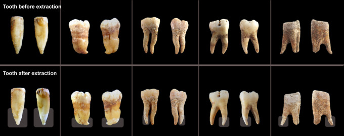
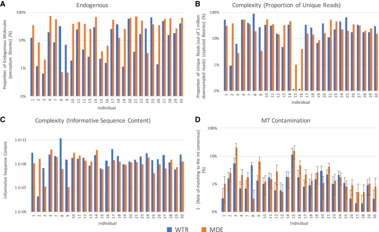
Ancient DNA sampling methods-although optimized for efficient DNA extraction-are destructive, relying on drilling or cutting and powdering (parts of) bones and teeth. As the field of ancient DNA has grown, so have concerns about the impact of destructive sampling of the skeletal remains from which ancient DNA is obtained. Due to a particularly high concentration of endogenous DNA, the cementum of tooth roots is often targeted for ancient DNA sampling, but destructive sampling methods of the cementum often result in the loss of at least one entire root. Here, we present a minimally destructive method for extracting ancient DNA from dental cementum present on the surface of tooth roots. This method does not require destructive drilling or grinding, and, following extraction, the tooth remains safe to handle and suitable for most morphological studies, as well as other biochemical studies, such as radiocarbon dating. We extracted and sequenced ancient DNA from 30 teeth (and nine corresponding petrous bones) using this minimally destructive extraction method in addition to a typical tooth sampling method. We find that the minimally destructive method can provide ancient DNA that is of comparable quality to extracts produced from teeth that have undergone destructive sampling processes. Further, we find that a rigorous cleaning of the tooth surface combining diluted bleach and UV light irradiation seems sufficient to minimize external contaminants usually removed through the physical removal of a superficial layer when sampling through regular powdering methods.


Figures

Figure 1.
Figure 1.
Examples of teeth before and after minimally destructive extraction. Teeth which have been sampled using this minimally destructive extraction protocol were photographed prior to (top) and ∼24 h after (bottom) extraction. Through the use of parafilm to protect regions of the tooth that are not targeted during sampling, such as the crown, sample degradation is primarily restricted to the lower portion of the targeted tooth roots, and the overall morphology of the tooth remains intact. The region targeted for sampling (i.e., not covered by parafilm) is indicated by a transparent box in the after images. Note that these are representative examples of the typical impact of sampling using this method on ancient teeth of high quality (two left-most teeth) or moderate quality (three right-most teeth). Data from these teeth are not reported in this study. For before and after images of the tooth roots upon which sequencing was done during this study, see Supplemental Figure S1.
Figure 2.
Figure 2.
Sample quality. A comparison of the quality of data produced by WTR (Whole Tooth Root) and MDE (Minimally Destructive Extraction) methods in samples that passed quality filtering. (A) The proportion of endogenous molecules in data obtained via shotgun sequencing. (B) The complexity of each sample, as measured by the proportion of unique reads out of 1,000,000 reads sequenced. Asterisks indicate that the total number of unique reads sequenced was below 1,000,000 for the specified sample, and therefore complexity estimates could not be generated. (C) The complexity of each sample, as measured by informative sequence content. (D) The rate of contamination is compared by considering the rate of matching to mitochondrial consensus sequence. Error bars indicate the 95% confidence interval. Only samples that passed quality screening are shown. Plots showing comparisons with samples generated using Method P are shown in Supplemental Figure S2.

References

    1. Behar DM, van Oven M, Rosset S, Metspalu M, Loogväli E-L, Silva NM, Kivisild T, Torroni A, Villems R. 2012. A “Copernican” reassessment of the human mitochondrial DNA tree from its root. Am J Hum Genet 90: 675–684. 10.1016/j.ajhg.2012.03.002 - DOI - PMC - PubMed
    1. Bell LS, Kayser M, Jones C. 2008. The mineralized osteocyte: a living fossil. Am J Phys Anthropol 137: 449–456. 10.1002/ajpa.20886 - DOI - PubMed
    1. Bolnick DA, Bonine HM, Mata-Míguez J, Kemp BM, Snow MH, LeBlanc SA. 2012. Nondestructive sampling of human skeletal remains yields ancient nuclear and mitochondrial DNA. Am J Phys Anthropol 147: 293–300. 10.1002/ajpa.21647 - DOI - PubMed
    1. Bosshardt DD, Selvig KA. 1997. Dental cementum: the dynamic tissue covering of the root. Periodontol 2000 13: 41–75. 10.1111/j.1600-0757.1997.tb00095.x - DOI - PubMed
    1. Carpenter ML, Buenrostro JD, Valdiosera C, Schroeder H, Allentoft ME, Sikora M, Rasmussen M, Gravel S, Guillén S, Nekhrizov G, et al. 2013. Pulling out the 1%: whole-genome capture for the targeted enrichment of ancient DNA sequencing libraries. Am J Hum Genet 93: 852–864. 10.1016/j.ajhg.2013.10.002 - DOI - PMC - PubMed

Publication types

LinkOut - more resources