. 2021 Feb 16;118(7):e2025070118.
doi: 10.1073/pnas.2025070118.

METTL3-dependent N6-methyladenosine RNA modification mediates the atherogenic inflammatory cascades in vascular endothelium

Affiliations

METTL3-dependent N6-methyladenosine RNA modification mediates the atherogenic inflammatory cascades in vascular endothelium

Chian-Shiu Chien et al. Proc Natl Acad Sci U S A. .

Abstract

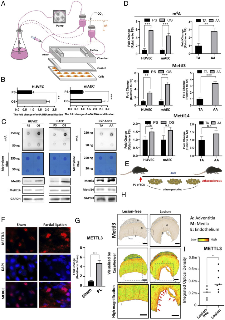
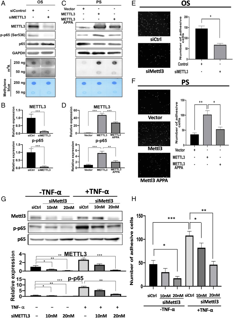
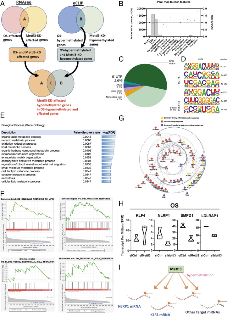
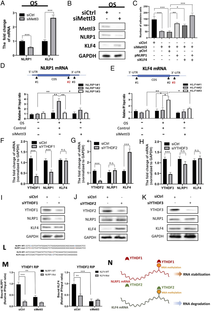
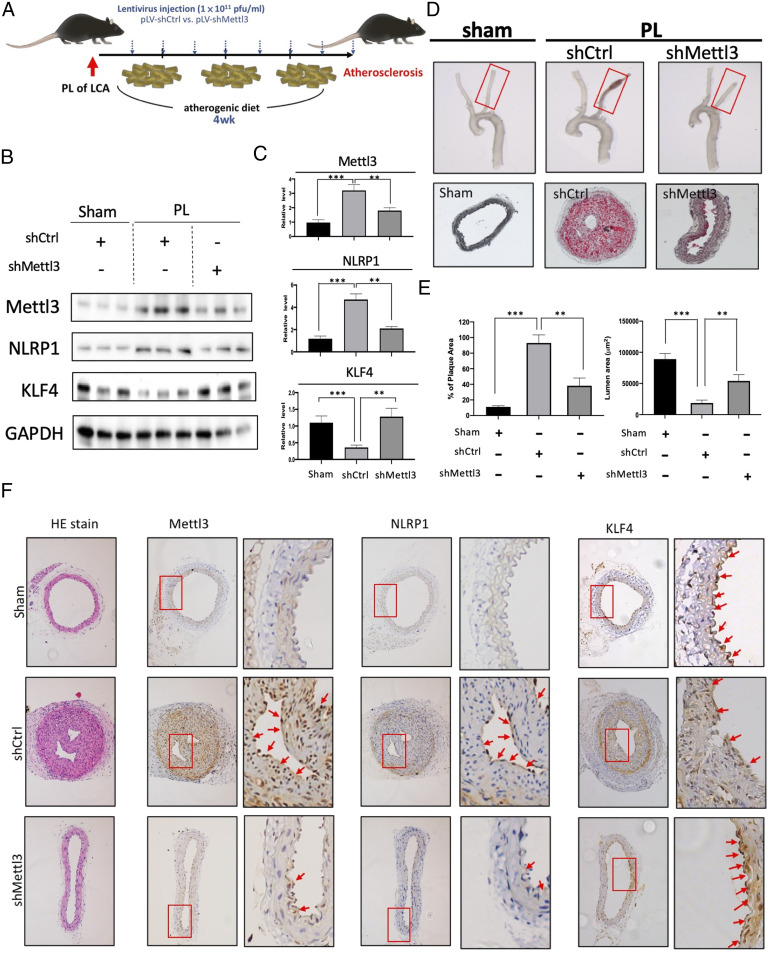
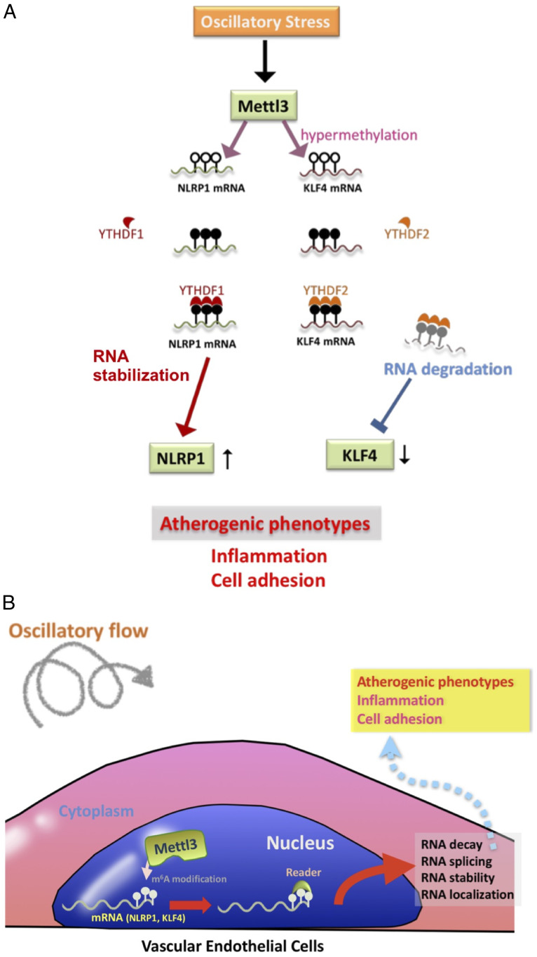
Atherosclerosis is characterized by the plaque formation that restricts intraarterial blood flow. The disturbed blood flow with the associated oscillatory stress (OS) at the arterial curvatures and branch points can trigger endothelial activation and is one of the risk factors of atherosclerosis. Many studies reported the mechanotransduction related to OS and atherogenesis; however, the transcriptional and posttranscriptional regulatory mechanisms of atherosclerosis remain unclear. Herein, we investigated the role of N6-methyladenosine (m6A) RNA methylation in mechanotransduction in endothelial cells (ECs) because of its important role in epitranscriptome regulation. We have identified m6A methyltransferase METTL3 as a responsive hub to hemodynamic forces and atherogenic stimuli in ECs. OS led to an up-regulation of METTL3 expression, accompanied by m6A RNA hypermethylation, increased NF-κB p65 Ser536 phosphorylation, and enhanced monocyte adhesion. Knockdown of METTL3 abrogated this OS-induced m6A RNA hypermethylation and other manifestations, while METTL3 overexpression led to changes resembling the OS effects. RNA-sequencing and m6A-enhanced cross-linking and immunoprecipitation (eCLIP) experiments revealed NLRP1 and KLF4 as two hemodynamics-related downstream targets of METTL3-mediated hypermethylation. The METTL3-mediated RNA hypermethylation up-regulated NLRP1 transcript and down-regulated KLF4 transcript through YTHDF1 and YTHDF2 m6A reader proteins, respectively. In the in vivo atherosclerosis model, partial ligation of the carotid artery led to plaque formation and up-regulation of METTL3 and NLRP1, with down-regulation of KLF4; knockdown of METTL3 via repetitive shRNA administration prevented the atherogenic process, NLRP3 up-regulation, and KLF4 down-regulation. Collectively, we have demonstrated that METTL3 serves a central role in the atherogenesis induced by OS and disturbed blood flow.

Keywords: METTL3; N6-methyladeosine RNA methylation; atherosclerosis; oscillatory flow; shear stress.


Conflict of interest statement

The authors declare no competing interest.

Figures

Fig. 1.
Fig. 1.
OS induces METTL3-dependent m6A methylation. (A) Scheme of the parallel-plate flow chamber for generating OS and PS at shear stresses of 0.5 ± 4 dyn/cm2 and 12 ± 4 dyn/cm2, respectively. (B) LCMS analysis of the content of m6A-modified mRNA in MAECs and HUVECs under PS or OS. Mean relative values are shown with SD error bars, **P < 0.05, ***P < 0.005. (C) Dot blot analysis of the contents of m6A-modified mRNA (Upper) and Western blot (Lower) of METTL3 and METTL14 protein expression in HUVECs and MAECs exposed to PS and OS, and in TA and AA from WT C57BL/6J mice. (D) Quantification of dot blot and Western blot. The mean values relative to PS or OS are shown with SD error bars, ***P < 0.005; NS, not significant. (E) Scheme for the induction of atherosclerosis in high-cholesterol atherogenic diet-fed ApoE−/− mice with partial ligation of left coronary artery. (F) En face immunofluorescence staining of METTL3 in the endothelial layer of PL LCA and sham control LCA. (G) Quantification showing mean fluorescence intensity values relative to sham control LCA with SD error bars, ***P < 0.005. (H, Top) Immunohistochemistry staining of the human paraffin-embedded tissue array of lesion-free aorta and aorta affected by atherosclerosis. (Middle) CaseViewer software was used to visualize METTL3-positive staining in these immunohistochemistry samples. The images were further zoomed in to show the endothelium with higher magnification. (I) The quantification showing METTL3 expression in the paraffin-embedded cross-sections of the human artery walls of the indicated samples. Quantification of METTL3 signal expressed as median and quartile integrated optical density (IOD).
Fig. 2.
Fig. 2.
METTL3 mediates proinflammatory effects in ECs. (A and B) Western blot (A) and its quantification expression (B) of METTL3, p65 and p-p65 (Upper), with dot blot analysis of the contents of m6A-modified RNA (Lower in A) in HUVECs treated with OS and METTL3 knockdown. (C and D) Western blot (C) and its quantification of expressions (D) of METTL3, p65 and p-p65 (Upper), dot blot analysis of the content of m6A-modified RNA (Lower in C) in HUVECs treated with PS and overexpression of METTL3 or its catalytic mutant METTL3 APPA. Monocyte adhesion assay: HUVECs treated with OS and METTL3 knockdown (E), and with PS and overexpression of METTL3 or its catalytic mutant METTL3 APPA (F). Quantification (Upper) and the representative fluorescent images of monocyte adhesion of five images are shown. SD: error bars, *P < 0.05, **P < 0.005. (G) Western blot (Upper) and quantification (Lower) of expressions of METTL3, p65, and p-p65 in HUVECs with METTL3 knockdown by siMETTL3 at 5 μg (#1) or 10 μg (#2) in the absence or presence of TNF-α stimulation. (Lower) Quantification of the blots with indicated treatment. (H) Monocyte adhesion assay performed on HUVECs subjected to METTL3 knockdown by siMETTL3 at 5 μg (#1) or 10 μg (#2) with or without TNF-α stimulation. Data shown as mean number of adherent monocytes from six images, SD error bars, *P < 0.05.
Fig. 3.
Fig. 3.
Identification of downstream m6A methylation sites of METTL3 in OS-treated HUVECs using NGS-based approaches and eCLIP analysis. (A) Venn diagrams showing the corresponding gene numbers of genes affected by OS and METTL3-knockdown (Upper Left) and the gene numbers of OS-hypermethylated genes and METTL3-knockdown hypomethylated genes (Lower). The overlap between OS-affected genes that were also affected by Mettl3-knockdown (label A) and OS-hypermethylated genes that were hypomethylated by Mettl3-knockdown (label B) represents the total m6A sites that were OS-affected and OS-hypomethylated by METTL3 knockdown (label C). (B) Bar charts showing the peaks that map to each feature and the ratio of shMettl3-treated HUVECs over shCtrl-treated HUVECs exposed to OS. (C) The pie chart depicts the relative frequency of peaks that map to each feature type, with a peak log2-fold enrichment ≥3 and P value ≤0.001. (D) HOMER identifies enriched motifs in CLIP-seq peaks. (E) The GO term enrichment analysis shows the total m6A sites affected and hypomethylated by METTL3 knockdown. Gene ratio is the number of genes annotated to the GO term in the input list of genes. Bg ratio is the number of genes annotated to that GO term in the entire background set. (F) GSEA of the total m6A sites that were affected and hypomethylated by METTL3 knockdown. (G) Identification of the network genes that are involved in the pathways related to total m6A sites that were affected and hypomethylated by METTL3 knockdown in OS-treated HUVECs. (H) RNA-seq profiles showing the transcript expressions of KLF4, NLRP1, SMPD1, and LDLRAP1. (I) Scheme illustrates that Mettl3 hypermethylates downstream mRNAs including KLF4, NLRP1, and other mRNAs in OS-exposed HUVECs.
Fig. 4.
Fig. 4.
Identification of m6A reader proteins that recognize METTL3-dependent methylation on NLRP1 and KLF4 mRNAs. (A) qRT-PCR analysis showing NLRP1 and KLF4 mRNA expressions in OS-treated HUVECs with or without Mettl3 knockdown. siCtrl, scrambled siRNA control. Mean values relative to siCtrl are shown with SD error bars, n = 3, ***P < 0.001. (B) Western blot showing NLRP1 and KLF4 proteins in OS-treated HUVECs with indicated treatment. (C) Monocyte adhesion assay performed on OS-treated HUVECs with or without Mettl3 knockdown, and with or without the concomitant NLRP1 overexpression (pNLRP1) and/or KLF4 knockdown (siKLF4). Data shown as means with SD error bars, n = 6, *P < 0.05, **P < 0.005, ***P < 0.001. (D and E) RNA-IP analysis showing the location of given m6A methylation sites in NLRP1 (D) or KLF4 mRNA (E) OS-treated HUVECs with indicated treatment. (F–H) qRT-PCR analysis showing the expression of YTHDF family members, NLRP1, and KLF4 mRNA in OS-treated HUVECs upon knockdown of YTHDF1 (F), YTHDF2 (G), and YTHDF3 (H). Data expressed as mean fold changes relative to control with SD error bars, n = 3, ***P < 0.001, n.s., not significant. (IK) Western blot of the expression of YTHDF family members, NLRP1, and KLF4 proteins upon the knockdown of YTHDF1 (I), YTHDF2 (J), or YTHDF3 (K) in OS-treated HUVECs. (L) Sequences of NLRP1 and KLF4 RNA fragments with either WT or mutated (mut) m6A sites. (M) RNA-IP assay showing the binding affinity between METTL3-hypermethylated target mRNAs and YTHDF family members with or without METTL3 knockdown. Synthesized RNA oligos of NLRP1 and KLF4 with either WT or mutated (Mut) m6A sites were introduced by electroporation, and the binding affinity between NLRP1 (Left) and YTHDF1 (Right), and between KLF4 and YTHDF2, were assessed. Data expressed as mean m6A enrichment relative to WT with SD error bars, n = 3, ***P < 0.001 (Student’s t test). (N) Scheme showing that YTHDF family members recognize METTL3-mediated m6A methylation on NLRP1 and KLF4 mRNAs under OS.
Fig. 5.
Fig. 5.
Mettl3 knockdown inhibits atherogenesis in vivo. (A) The experimental design for Mettl3 gene knockdown expression in the partial ligation (PL)-induced high-cholesterol atherogenic diet-fed ApoE−/− mouse model of atherosclerosis. Atherosclerotic lesions were induced by PL over 4 wk. Lentivirus encoding control shRNA (pLV-shCtrl) or shRNA targeting Mettl3 (pLV-shMettl3) was repetitively administered twice per week. (B) Western blot and its quantification (C) showing the expression of METTL3, NLRP1, and KLF4 proteins in the arterial endothelium from mice with indicated treatments. (D) Gross necropsy (Upper) of LCAs from ApoE−/− mice with indicated treatments. Plaque formation and the changes in lumen size (Center) were evaluated using Oil Red O staining. (E) The formation of Oil Red O-stained plaques and quantitation of lumen area. (F) Hematoxylin and eosin staining and immunohistochemistry analysis of METTL3 and NLRP1 proteins in the cross-sections from sham-operated LCA and PL LCA with indicated treatments. Boxes highlighted the expression patterns of METTL3, NLRP1, and KLF4 in the endothelium from LCA.
Fig. 6.
Fig. 6.
Scheme illustrating the role of METTL3-dependent m6A RNA modifications in OS-induced proatherogenic events. The working models of METTL3-dependent m6A RNA modifications in OS-induced proatherogenic events are shown. (A) OS up-regulates METTL3 and induces a METTL3-dependent m6A RNA hypermethylation in downstream mRNA targets, e.g., NLRP1 and KLF4. For NLRP1 mRNA, the METTL3-mediated hypermethylation is recognized by YTHDF1, leading to enhanced translation of NLRP1. For KLF4, YTHDF2 recognizes the methylation sites and further lead to the degradation of KLF4 mRNA. (B) Overall, m6A RNA modifications may induce RNA decay, splicing, stability, and their localization; further modulate downstream protein expression; and eventually contribute to proatherogenic events.

References

    1. Chiu J. J., Chien S., Effects of disturbed flow on vascular endothelium: Pathophysiological basis and clinical perspectives. Physiol. Rev. 91, 327–387 (2011). - PMC - PubMed
    1. Ajami N. E., et al. , Systems biology analysis of longitudinal functional response of endothelial cells to shear stress. Proc. Natl. Acad. Sci. U.S.A. 114, 10990–10995 (2017). - PMC - PubMed
    1. Neth P., Nazari-Jahantigh M., Schober A., Weber C., MicroRNAs in flow-dependent vascular remodelling. Cardiovasc. Res. 99, 294–303 (2013). - PubMed
    1. Geula S., et al. , Stem cells. m6A mRNA methylation facilitates resolution of naïve pluripotency toward differentiation. Science 347, 1002–1006 (2015). - PubMed
    1. Dunn J., Simmons R., Thabet S., Jo H., The role of epigenetics in the endothelial cell shear stress response and atherosclerosis. Int. J. Biochem. Cell Biol. 67, 167–176 (2015). - PMC - PubMed

Publication types

MeSH terms