

Comment
doi: 10.1038/s41467-021-21074-x.
Sulfisoxazole does not inhibit the secretion of small extracellular vesicles
Affiliations
- PMID: 33579909
- PMCID: PMC7881022
- DOI: 10.1038/s41467-021-21074-x
Item in Clipboard
Comment
Sulfisoxazole does not inhibit the secretion of small extracellular vesicles
Nat Commun.
.
No abstract available
Conflict of interest statement
The authors declare no competing interests.
Figures
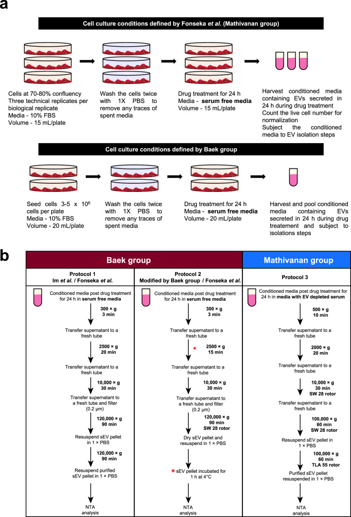
a Schematic representation of cell culture and drug treatment approaches. The cells were seeded and allowed to grow to 70–80% confluency in media supplemented with FCS. Upon attaining desired confluency, cells were washed with 1× PBS and subjected to drug treatment for 24 h in serum-free media. At treatment endpoint, conditioned media was harvested and subjected to sEV isolation. Live cell number was determined to normalise particles released to cell number. Similarly, Baek group seeded cells in a pre-defined range and treated them with drug in serum-free media. b Protocol 1, schematic flow diagram depicting the methodology of sEV isolation and analyses as defined by Baek group in the initial publication (Im et al.) and followed by Fonseka et al. (this study). Protocol 2, schematic flow diagram depicts the methodology of sEV isolation and analyses as reported by Baek group when approached. The variations in the methodology from the original manuscript (Im et al.) is marked (*). Protocol 2 was also adapted by Fonseka et al. to include the variations (*) suggested by Baek group. Protocol 3, standardized and optimized method of sEV isolation in EV depleted serum used in Mathivanan laboratory was also used to collect sEVs post treatment with SFX.
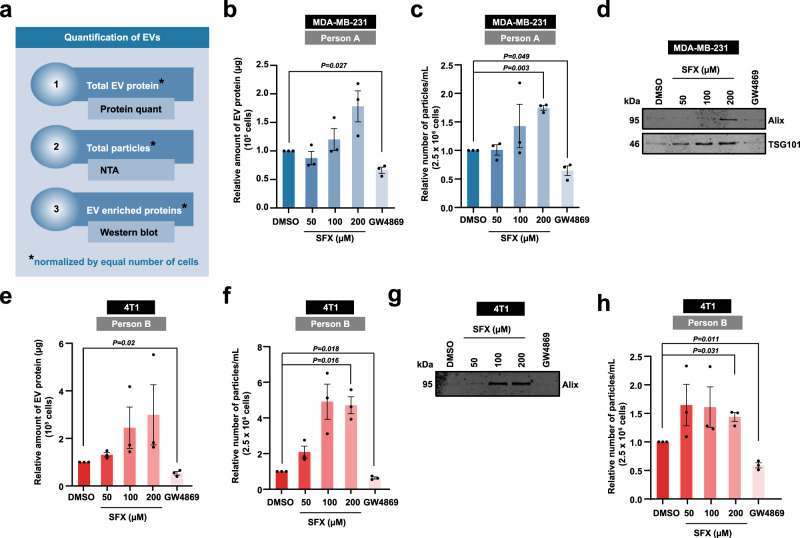
a Schematic of EV quantification by three different methods. Firstly, the total EV protein amount was quantified and normalised to equal number of live cells. Secondly, nanoparticle tracking analysis (NTA) was performed to quantify the total number of particles normalised to equal number of live cells. Lastly, Western blot analysis of EV samples obtained from equal number of live cells was performed for EV enriched proteins. b Relative amount of EV protein normalised to 105 MDA-MB-231 cells is shown. c Relative number of particles normalised to 2.5 × 10⁶ MDA-MB-231 cells is depicted. d Western blot analysis of EV enriched proteins Alix and TSG101 in EV samples obtained from 15 × 106 MDA-MB-231 cells. Representative image shown, n = 3 biologically independent experiments. e Relative amount of EV protein normalised to 105 4T1 cells is shown. f Relative number of particles normalised to 2.5 × 10⁶ 4T1 cells is depicted. g Western blot analysis of EV enriched proteins Alix in EV samples obtained from 15 × 106 4T1 cells. Representative image shown, n = 3 biologically independent experiments. h Relative number of particles normalised to 2.5 × 10⁶ 4T1 cells is depicted. All data are represented as mean ± s.e.m. n = 3 biologically independent experiments, statistical significance was determined by paired two-tailed t-test. EVs were isolated by protocol 1 (b–g) and 2 (h). Full uncropped images for western blotting is provided in Supplementary Fig. 1.
Comment in
-
Reply to 'Sulfisoxazole does not inhibit the secretion of small extracellular vesicles'.Nat Commun. 2021 Feb 12;12(1):976. doi: 10.1038/s41467-021-21075-w. Nat Commun. 2021. PMID: 33579906 Free PMC article. No abstract available.
Comment on
-
Sulfisoxazole inhibits the secretion of small extracellular vesicles by targeting the endothelin receptor A.Nat Commun. 2019 Mar 27;10(1):1387. doi: 10.1038/s41467-019-09387-4. Nat Commun. 2019. PMID: 30918259 Free PMC article.
References
-
- Thery C, et al. Minimal information for studies of extracellular vesicles 2018 (MISEV2018): a position statement of the International Society for Extracellular Vesicles and update of the MISEV2014 guidelines. J. Extracell. Vesicles. 2018;7:1535750. doi: 10.1080/20013078.2018.1535750. - DOI - PMC - PubMed
Publication types
MeSH terms
Substances
LinkOut - more resources
Full Text Sources
Other Literature Sources
