. 2021 Feb 12;12(1):1013.
doi: 10.1038/s41467-021-21129-z.

AA amyloid fibrils from diseased tissue are structurally different from in vitro formed SAA fibrils

Affiliations

AA amyloid fibrils from diseased tissue are structurally different from in vitro formed SAA fibrils

Akanksha Bansal et al. Nat Commun. .

Abstract

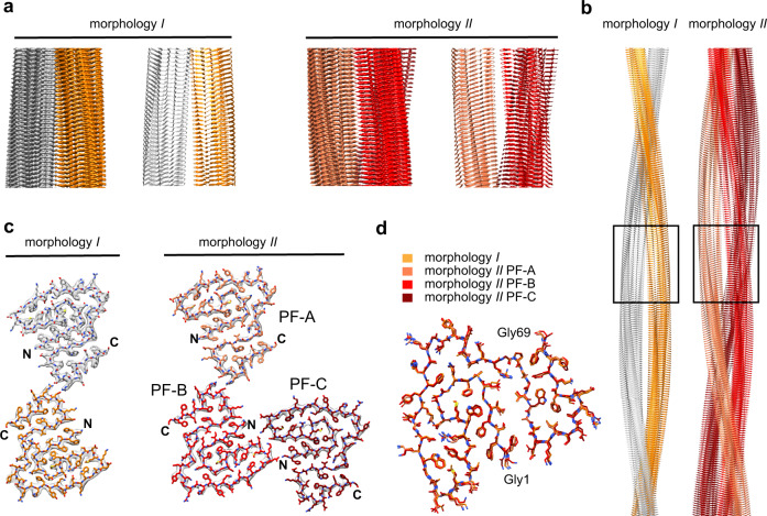
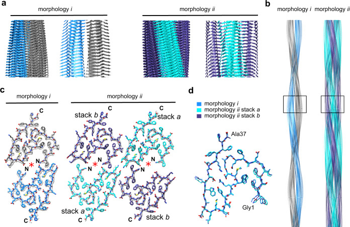
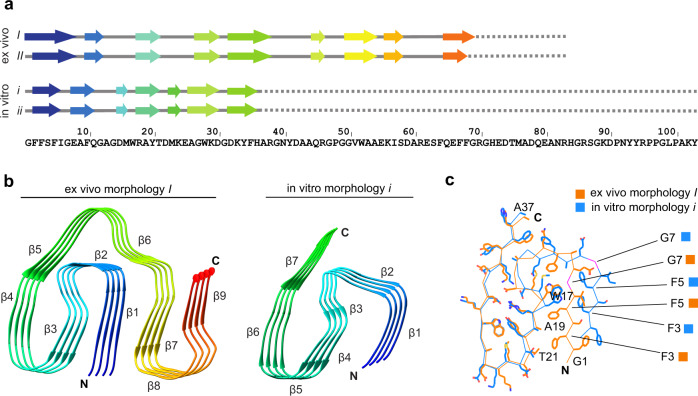
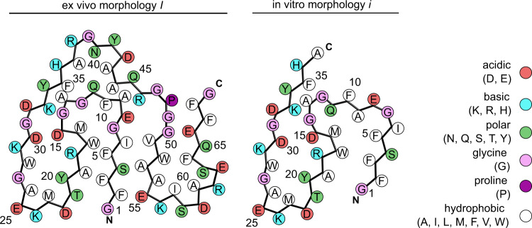
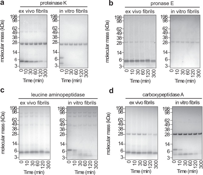
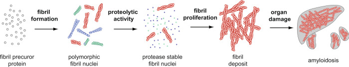
Systemic AA amyloidosis is a world-wide occurring protein misfolding disease of humans and animals. It arises from the formation of amyloid fibrils from serum amyloid A (SAA) protein. Using cryo electron microscopy we here show that amyloid fibrils which were purified from AA amyloidotic mice are structurally different from fibrils formed from recombinant SAA protein in vitro. Ex vivo amyloid fibrils consist of fibril proteins that contain more residues within their ordered parts and possess a higher β-sheet content than in vitro fibril proteins. They are also more resistant to proteolysis than their in vitro formed counterparts. These data suggest that pathogenic amyloid fibrils may originate from proteolytic selection, allowing specific fibril morphologies to proliferate and to cause damage to the surrounding tissue.


Conflict of interest statement

The authors declare no competing interests.

Figures

Fig. 1
Fig. 1. Cryo-EM structures of AA amyloid fibrils purified from diseased tissue.
a Side views of fibril morphologies I and II: corresponding segments of the 3D maps and models, shown as ribbon diagrams. b Models of fibrils in side view. The segment shown in a is boxed. c Cross-sectional layers of fibril morphologies I and II. The 3D maps (gray surfaces) are superimposed with the models, shown as sticks. d Alignment of the fibril proteins of morphology I and II (PF-A to PF-C) as indicated in the figure. The data of morphology I is taken from a previously deposited data (PDB 6DSO: https://doi.org/10.2210/pdb6dso/pdb).
Fig. 2
Fig. 2. Cryo-EM structures of amyloid fibrils from recombinant SAA1.1 protein in vitro.
a Side views of fibril morphologies i and ii: corresponding sections of the 3D maps and models, shown as ribbon diagrams. b Models of fibrils in side view. The segment shown in a is boxed. c Cross-sectional layers of fibril morphologies i and ii. The 3D maps (gray surfaces) are superimposed with the models, shown as sticks. Red asterisk: cavity of the structure. d Alignment of the fibril proteins of morphology i and ii (stacks a and b) as indicated in the figure.
Fig. 3
Fig. 3. Fibril protein conformation in in vitro and ex vivo fibrils.
a Sequence of SAA1.1 indicating the location of the β-strands. b Ribbon diagrams of fibril protein stacks (ex vivo fibril morphology I and in vitro fibril morphology i) colored in rainbow palette. c Alignment of residues 1–37 of the ex vivo (orange) and in vitro fibril protein structure (blue).
Fig. 4
Fig. 4. Schematic view of fibril protein packing in ex vivo fibril morphology I and in vitro fibril morphology i.
Comparison of complementary fibril protein packing in ex vivo fibril morphology I and in in vitro fibril morphology i.
Fig. 5
Fig. 5. Stability of the fibrils against proteolysis.
Coomassie-stained denaturing protein gels of ex vivo AA amyloid fibrils and in vitro formed SAA1.1 fibrils that were incubated with different proteases for up to 300 min: proteinase K (a, n = 3) and three other proteases (pronase E (b), leucine aminopeptidase (c) and carboxypeptidase A (d), n = 1). Bands above 14 kDa originate from the proteases. Due to the harsh proteolytic conditions used in this experiment there is discernible degradation of in vitro fibrils during sample workup for electrophoresis (0 min), specifically with proteinase K and pronase E.
Fig. 6
Fig. 6. Schematic representation of the proteolytic selection mechanism.
Proliferation and subsequent organ deposition of proteolytically selected pathogenic fibril morphologies. The protease unstable morphologies have been depicted in blue and green while the protease stable morphology is shown in red.

References

    1. Dobson CM. The structural basis of protein folding and its links with human disease. Philos. Trans. R. Soc. Lond. B. 2001;356:133–145. doi: 10.1098/rstb.2000.0758. - DOI - PMC - PubMed
    1. Eisenberg D, Jucker M. The amyloid state of proteins in human diseases. Cell. 2012;148:1188–1203. doi: 10.1016/j.cell.2012.02.022. - DOI - PMC - PubMed
    1. Benson MD, et al. Amyloid nomenclature 2018: recommendations by the International Society of Amyloidosis (ISA) nomenclature committee. Amyloid. 2018;25:215–219. doi: 10.1080/13506129.2018.1549825. - DOI - PubMed
    1. Annamalai K, et al. Common fibril structures imply systemically conserved protein misfolding pathways in vivo. Angew. Chem. Int. Ed. Engl. 2017;56:7510–7514. doi: 10.1002/anie.201701761. - DOI - PubMed
    1. Radamaker, L. et al. Cryo-EM reveals structural breaks in a patient-derived amyloid fibril from systemic AL amyloidosis. Preprint at 10.1101/2020.10.12.332569 (2020). - PMC - PubMed

Publication types