. 2021 Feb 12;5(1):10.
doi: 10.1038/s41698-021-00147-6.

PD-L1 immunohistochemistry for canine cancers and clinical benefit of anti-PD-L1 antibody in dogs with pulmonary metastatic oral malignant melanoma

Affiliations

PD-L1 immunohistochemistry for canine cancers and clinical benefit of anti-PD-L1 antibody in dogs with pulmonary metastatic oral malignant melanoma

Naoya Maekawa et al. NPJ Precis Oncol. .

Abstract

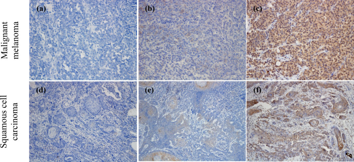
Immunotherapy targeting programmed cell death 1 (PD-1) and PD-ligand 1 (PD-L1) represents promising treatments for human cancers. Our previous studies demonstrated PD-L1 overexpression in some canine cancers, and suggested the therapeutic potential of a canine chimeric anti-PD-L1 monoclonal antibody (c4G12). However, such evidence is scarce, limiting the clinical application in dogs. In the present report, canine PD-L1 expression was assessed in various cancer types, using a new anti-PD-L1 mAb, 6C11-3A11, and the safety and efficacy of c4G12 were explored in 29 dogs with pulmonary metastatic oral malignant melanoma (OMM). PD-L1 expression was detected in most canine malignant cancers including OMM, and survival was significantly longer in the c4G12 treatment group (median 143 days) when compared to a historical control group (n = 15, median 54 days). In dogs with measurable disease (n = 13), one dog (7.7%) experienced a complete response. Treatment-related adverse events of any grade were observed in 15 dogs (51.7%). Here we show that PD-L1 is a promising target for cancer immunotherapy in dogs, and dogs could be a useful large animal model for human cancer research.


Conflict of interest statement

Authors S.Y., K.Y., and M.T. were employed by Fuso Pharmaceutical Industries, Ltd. S. Konnai, K.O., S.M., Y.S., C.N., T.O., N.M., S.T., and Y. Kagawa are the authors of patent applications covering materials and techniques described in this paper (PCT/JP2017/029055, PCT/JP2018/11895). All other authors declare no competing interests.

Figures

Fig. 1
Fig. 1. Representative PD-L1 immunohistochemistry results with 6C11-3A11 for each tumor proportion score (TPS).
The proportion of PD-L1–expressing tumor cells was scored according to the percent of stained viable tumor cells (see “Methods”). Representative IHC results for TPS < 1% (a, d), 1–49% (b, e), and ≥50% (c, f) are shown. Original magnification, ×200 (ac) or ×100 (df).
Fig. 2
Fig. 2. Antitumor efficacies of c4G12 in dogs with oral malignant melanoma.
a Representative tumor response in dogs with measurable disease. The dog (Pomeranian, male, 12-year-old, #10 in Supplementary Tables 2, 3) received c4G12 at a dose of 5 mg/kg every 2 weeks. Oral recurrent tumor (upper panel) and pulmonary metastatic lesions (lower panel) responded to treatment at week 7. b, c Representative tumor response in dogs with non-measurable disease. b The dog (Pekingese, castrated male, 8-year-old, #12) received c4G12 at a dose of 5 mg/kg every 2 weeks. Pulmonary metastatic lesions were confirmed at week 4. Subsequently, the lesions responded to treatment at week 12. c The dog (Beagle, spayed female, 11-year-old, #5) received c4G12 at a dose of 5 mg/kg every 2 weeks until week 12 and thereafter the dose was changed to 2 mg/kg. A metastatic lesion in the lung (upper panel) disappeared at week 6. In contrast, the other lesion (lower panel) showed slight increase in tumor size at week 6, followed by complete regression at week 18 (an immune-related pattern of response). Contrast-enhanced and matched transverse CT images are shown. Arrow heads indicate tumor lesions.
Fig. 3
Fig. 3. Survival of dogs treated with c4G12.
a Comparison of survival between c4G12 treatment group (n = 29) and historical control group (n = 15). Survival (days) was defined as time from confirmation of pulmonary metastasis to death. For subgroup analysis, the treatment group was dichotomized according to (b) the use of previous/concomitant radiation, (c) plasma C reactive protein (CRP) levels (cutoff 2.55 mg/dL), or (d) the lymphocyte-to-monocyte ratio (LMR, cutoff 1.41), and overall survival was compared between the subgroups. Dogs that received radiation therapy concomitantly or ≤8 weeks before the initiation of antibody treatment were classified into radiation (+) subgroup. Marks on the line indicate censored data. Statistical analysis was performed using the log-rank test.

References

    1. Iwai Y, Hamanishi J, Chamoto K, Honjo T. Cancer immunotherapies targeting the PD-1 signaling pathway. J. Biomed. Sci. 2017;24:26. doi: 10.1186/s12929-017-0329-9. - DOI - PMC - PubMed
    1. Keir ME, Butte MJ, Freeman GJ, Sharpe AH. PD-1 and its ligands in tolerance and immunity. Annu. Rev. Immunol. 2008;26:677–704. doi: 10.1146/annurev.immunol.26.021607.090331. - DOI - PMC - PubMed
    1. Chen L, Flies DB. Molecular mechanisms of T cell co-stimulation and co-inhibition. Nat. Rev. Immunol. 2013;13:227–242. doi: 10.1038/nri3405. - DOI - PMC - PubMed
    1. Dong H, et al. Tumor-associated B7-H1 promotes T-cell apoptosis: a potential mechanism of immune evasion. Nat. Med. 2002;8:793–800. doi: 10.1038/nm730. - DOI - PubMed
    1. Sznol M, Chen L. Antagonist antibodies to PD-1 and B7-H1 (PD-L1) in the treatment of advanced human cancer. Clin. Cancer Res. 2013;19:1021–1034. doi: 10.1158/1078-0432.CCR-12-2063. - DOI - PMC - PubMed