Review
. 2021 Apr;99(4):531-553.
doi: 10.1007/s00109-020-02028-0. Epub 2021 Feb 13.

Organoids of the female reproductive tract

Affiliations
Review

Organoids of the female reproductive tract

Cindrilla Chumduri et al. J Mol Med (Berl). 2021 Apr.

Abstract

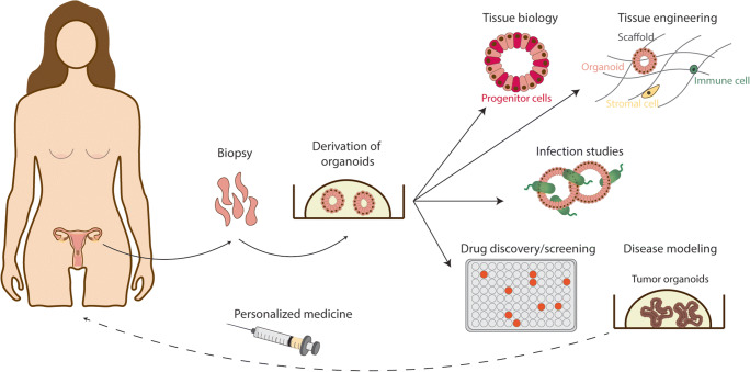
Healthy functioning of the female reproductive tract (FRT) depends on balanced and dynamic regulation by hormones during the menstrual cycle, pregnancy and childbirth. The mucosal epithelial lining of different regions of the FRT-ovaries, fallopian tubes, uterus, cervix and vagina-facilitates the selective transport of gametes and successful transfer of the zygote to the uterus where it implants and pregnancy takes place. It also prevents pathogen entry. Recent developments in three-dimensional (3D) organoid systems from the FRT now provide crucial experimental models that recapitulate the cellular heterogeneity and physiological, anatomical and functional properties of the organ in vitro. In this review, we summarise the state of the art on organoids generated from different regions of the FRT. We discuss the potential applications of these powerful in vitro models to study normal physiology, fertility, infections, diseases, drug discovery and personalised medicine.

Keywords: Cancers; Female reproductive tract; Fertility; Infection; Organoids; Pregnancy; Reproductive health.


Conflict of interest statement

The authors declare that they have no conflict of interest.

Figures

Fig. 1
Fig. 1
Anatomy of the human and mouse FRT. In humans, the FRT consists of ovaries, fallopian tubes, uterus (endometrium and myometrium), cervix (endocervix and ectocervix) and vagina. There are some anatomical differences between the two species, as mice have a bicornuate uterus (uterine horns) and oviducts are much less pronounced in proportion to the rest of the reproductive tract
Fig. 2
Fig. 2
The menstrual cycle. The pituitary and ovarian hormones regulating the menstrual cycle and the morphological changes occurring at the ovarian surface epithelium (OSE) and endometrium are depicted. The menstrual phase is divided into menstrual, proliferative and secretory phases. During the proliferative phase, follicle-stimulating hormone (FSH) promotes the growth of ovarian follicles. This results in rising levels of oestrogen (E2) produced by the follicles which then drives the proliferation of the functional layer of the endometrium. Mid-cycle, a peak in E2 results in a surge of luteinizing hormone (LH) and the release of the oocyte (ovulation). This marks the start of the secretory phase, dominated by progesterone (P4), which drives the differentiation (decidualisation) of the endometrium to prepare for implantation. In the absence of implantation, P4 levels drop triggering menstruation, the shedding of the functional layer, to restart the cycle. The basal layer is not shed
Fig. 3
Fig. 3
The epithelia of the human FRT. The types of epithelia covering the different regions of the human FRT are illustrated. For each tissue the major cell types are shown, with examples of typical markers they express. Markers with an asterisk are hormonally regulated
Fig. 4
Fig. 4
Organoids of normal and diseased tissues of the human FRT. Organoid models derived from normal and pathological tissues are illustrated. The different cell types that are present in the tissue epithelia and the organoids are shown as columnar (non-ciliated), secretory, ciliated, cuboidal and squamous. The organoids recapitulate cellular heterogeneity, genetic signature and key functions of the tissue of origin
Fig. 5
Fig. 5
Future applications of organoids of the FRT. Organoids of the FRT are derived from biopsies and can be used to study tissue biology, cell-cell interactions, host-pathogen interactions and disease. Tissue engineering methods combine organoids with other cell types to develop more tissue-like models that include non-epithelial cells. Organoids derived from pathological tissues are useful for drug screening for personalised medicine

Similar articles

Cited by

References

    1. Kobayashi A, Behringer RR. Developmental genetics of the female reproductive tract in mammals. Nat Rev Genet. 2003;4:969–980. - PubMed
    1. Christensen A, Bentley GE, Cabrera R, et al. Hormonal regulation of female reproduction. Horm Metab Res. 2012;44:587–591. - PMC - PubMed
    1. Burney RO, Giudice LC (2012) Pathogenesis and pathophysiology of endometriosis. Fertil Steril 98. 10.1016/j.fertnstert.2012.06.029 - PMC - PubMed
    1. Bagnoli M, Shi TY, Gourley C et al (2019) Gynecological cancers translational, research implementation, and harmonization: Gynecologic Cancer InterGroup Consensus and Still Open Questions. Cells 8. 10.3390/cells8030200 - PMC - PubMed
    1. Onstad MA, Schmandt RE, Lu KH. Addressing the role of obesity in endometrial cancer risk, prevention, and treatment. J Clin Oncol. 2016;34:4225–4230. - PMC - PubMed

Publication types

MeSH terms

Substances

LinkOut - more resources