Review
. 2021 Apr:60:34-43.
doi: 10.1016/j.mib.2021.01.015. Epub 2021 Feb 10.

Resource sharing between central metabolism and cell envelope synthesis

Affiliations
Review

Resource sharing between central metabolism and cell envelope synthesis

Ankita J Sachla et al. Curr Opin Microbiol. 2021 Apr.

Abstract

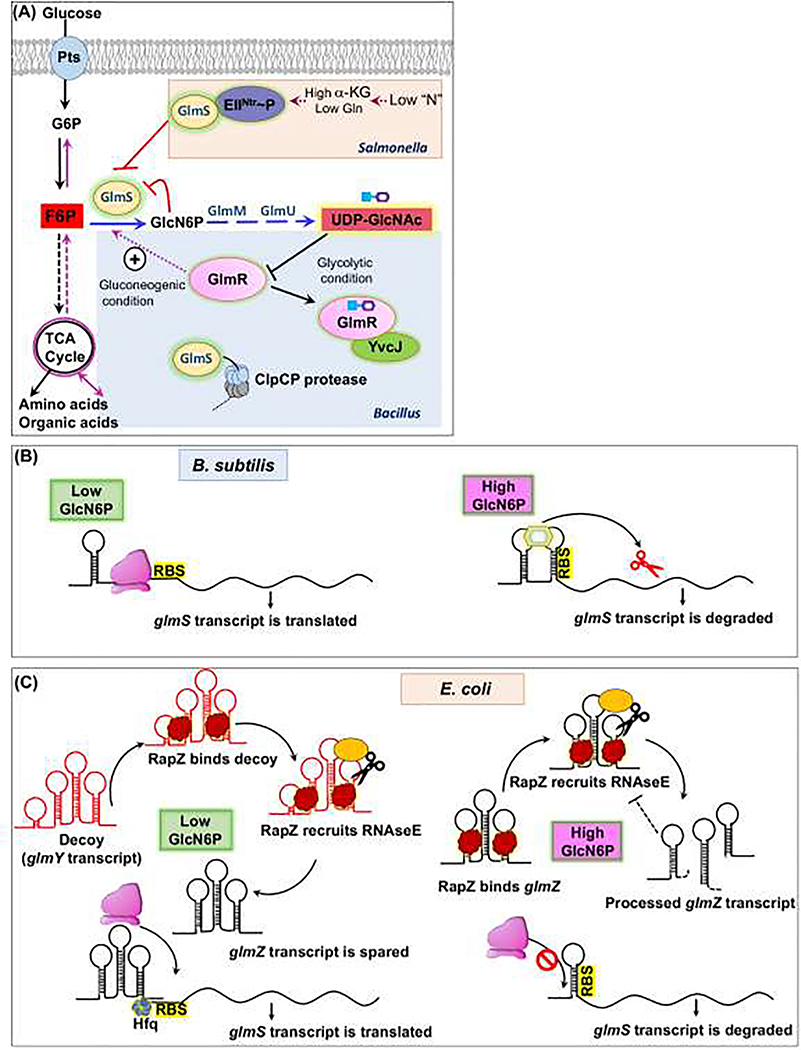
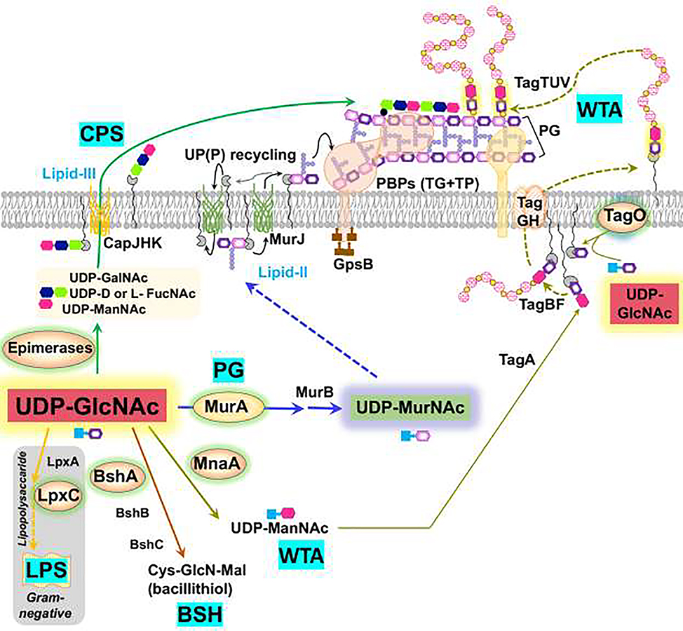
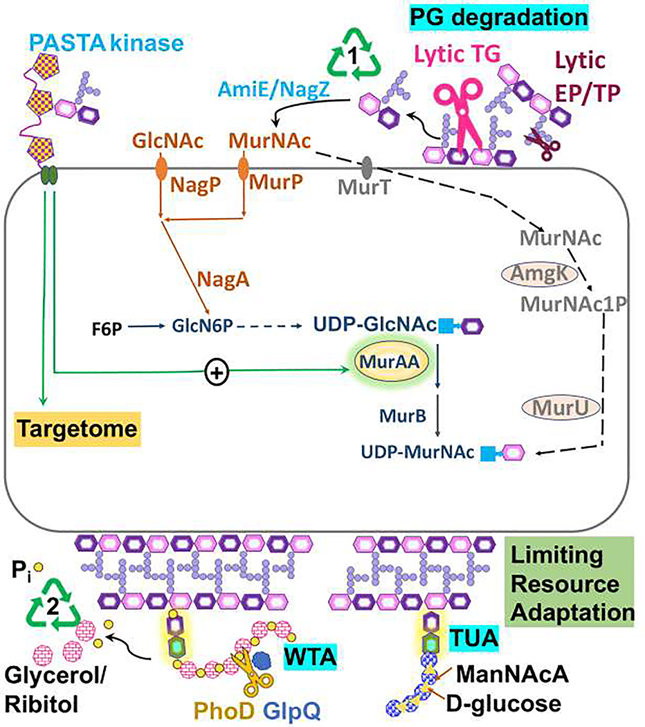
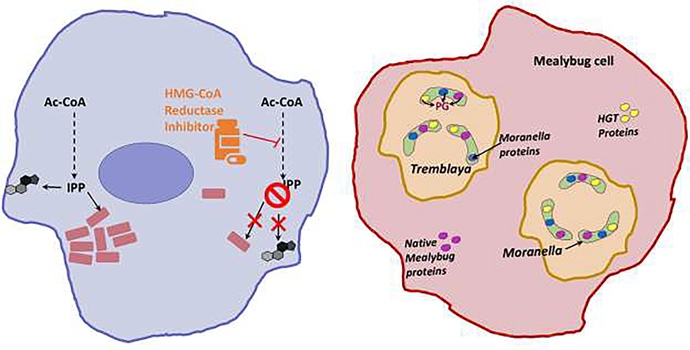
Synthesis of the bacterial cell envelope requires a regulated partitioning of resources from central metabolism. Here, we consider the key metabolic junctions that provide the precursors needed to assemble the cell envelope. Peptidoglycan synthesis requires redirection of a glycolytic intermediate, fructose-6-phosphate, into aminosugar biosynthesis by the highly regulated branchpoint enzyme GlmS. MurA directs the downstream product, UDP-GlcNAc, specifically into peptidoglycan synthesis. Other shared resources required for cell envelope synthesis include the isoprenoid carrier lipid undecaprenyl phosphate and amino acids required for peptidoglycan cross-bridges. Assembly of the envelope requires a sharing of limited resources between competing cellular pathways and may additionally benefit from scavenging of metabolites released from neighboring cells or the formation of symbiotic relationships with a host.


Conflict of interest statement

Conflict of interest statement.

Nothing declared.

Figures

Figure 1
Figure 1. Regulation of the GlmS branchpoint enzyme.
A) Post-translational regulatory mechanisms. Glucose (Glc) is imported through a PTS and enters glycolysis where the branchpoint metabolite fructose-6-phosphate (F6P) is partitioned to aminosugar biosynthesis by GlmS. In Salmonella Typhimurium, GlmS is inhibited by the phosphorylated EII protein of the PTSNTR system under conditions of nitrogen deficiency (sensed by a high α-ketoglutarate to glutamine ratio). In B. subtilis, GlmS is stimulated the regulator GlmR(YvcK), and this activation is inhibited by binding to the next key branchpoint metabolite UDP-GlcNAc (UDP=blue square; GlcNAc is purple hexagon), GlmR:UDP-GlcNAc binds to YvcJ, which reduced GlmS activity/ GlmS is also subject to ClpCP-dependent proteolysis. (B) Feedback regulation of GlmS translation. In B. subtilis, the glmS transcript includes a 5’-regulatory ribozyme that binds GlcN6P, resulting in cleavage and inactivation of the mRNA. (C) Translation of the E. coli glmS mRNA is activated for recognition by the ribosome (pink) when bound to the regulatory RNA glmZ. However, when GlcN6P is high the glmZ transcript is degraded by RNase E, which is recruited by RapZ. When GlcN6P is low, RapZ triggers expression of a decoy transcript (GlmY) that sequesters RapZ, and the GlmZ sRNA is spared to activate glmS translation.
Figure 2.
Figure 2.. UDP-GlcNAc is a key branchpoint metabolite.
UDP-GlcNAc (red) is a shared precursor directed to peptidoglycan (PG) synthesis by MurA. PG synthesis relies on a UP-linked disaccharide-pentapeptide (lipid II), which is exported by MurJ so that penicillin-binding proteins (PBPs) and the Rod complex (elongasome) can catalyze the transglycosylation (TG) and transpeptidation (TP) reactions for PG synthesis. In Gram-positive bacteria, UDP-GlcNAc is additionally directed to wall teichoic acid (WTA) synthesis by MnaA (for UDP-ManNAc synthesis) and TagO (which couples GlcNAc to UP). WTA polymers are synthesized in the cytosol, flipped across the membrane by the TagGH complex, and covalently linked to PG (TagTUV enzymes). UDP-GlcNAc is also required for bacillithiol (BSH) synthesis, CPS synthesis and, in Gram-negative bacteria, for LPS biosynthesis. Branchpoint enzymes are highlighted with a yellow background.
Figure 3.
Figure 3.. Degradation and recycling of the cell envelope.
Cell growth is accompanied by release of muropeptides generated by lytic transglycosylases (lytic TG) and endopeptidases/transpeptidases (lytic EP.TP) (1), which may be recycled. Recycling pathways involve degradation to release GlcNAc and MurNAc sugars and amino acids. Gram-negative bacteria may import muropeptodes and recycle intracellularly (not shown). In B. subtilis (orange), MurNAc and GlcNAc, are imported by PTS transporters MurP and NagP. The oral pathogen Tannerella forsythia is auxotrophic for MurNAc, which is imported by MurT and processed to generate UDPMurNAc (gray). Muropeptides can also activate some PASTA kinases, which phosphorylate many target proteins (targetome). In some systems, this can regulate the PG branchpoint enzyme MurA. (2) During phosphate limitation, Gram-positive bacteria may recycle WTA (glycerol-P copolymer) aided by the GlpQ and PhoD proteins. Secreted teichoicases may also cleave WTA off of nearby cells to scavenge phosphate. Phosphate limitation in B. subtilis triggers a switch from synthesis of a glycerol-P-based WTA to a functionally similar, but phosphate-free teichuronic acid polymer.
Figure 4.
Figure 4.. Resource sharing beyond kin.
Left: Rickettsia (pink rectangles) obtains isoprenoid precursors from its host. The inhibition of host HMG-CoA reductase restricts this metabolic crossfeeding and ultimately affects Rickettsia survival. Right: Moranella is snugly housed in Tremblaya, which in turn lives as an endosymbiont in its insect host, the mealybug Planoccous citri cells. These nested cells share the burden of PG synthesis for the diminutive Moranella cell. The initial enzymes for PG biosynthesis are imported from the Mealybug and include both housekeeping enzymes (Glm-S,U, and M-depicted in pink) and enzymes acquired by horizontal gene transfer from Bacteria (MurA-F, DdlB, DapF, MltB, AmiD- depicted in yellow). The cytosolic PG intermediate synthesized by these imported enzymes can then be assembled by later stage enzymes retained by Moranella (MraY-Pbp’s- depicted in blue).

References

    1. Chubukov V, Gerosa L, Kochanowski K, Sauer U: Coordination of microbial metabolism. Nat Rev Microbiol 2014, 12:327–340. - PubMed
    1. Monk JM, Lloyd CJ, Brunk E, Mih N, Sastry A, King Z, Takeuchi R, Nomura W, Zhang Z, Mori H, et al.: iML1515, a knowledgebase that computes Escherichia coli traits. Nat Biotechnol 2017, 35:904–908. - PMC - PubMed
    1. Reith J, Mayer C: Peptidoglycan turnover and recycling in Gram-positive bacteria. Appl Microbiol Biotechnol 2011, 92:1–11. - PubMed
    1. Kawai Y, Mercier R, Mickiewicz K, Serafini A, Sorio de Carvalho LP, Errington J: Crucial role for central carbon metabolism in the bacterial L-form switch and killing by beta-lactam antibiotics. Nat Microbiol 2019, 4:1716–1726.

      • Blocking PG synthesis can trigger the emergence of L-forms, bacteria that lack a cell wall. However, the resultant increase in flux of carbon into lower glycolysis can ultimately the electron transfer chain triggers oxidative stress.

    1. Yoo W, Yoon H, Seok YJ, Lee CR, Lee HH, Ryu S: Fine-tuning of amino sugar homeostasis by EIIA(Ntr) in Salmonella Typhimurium. Sci Rep 2016, 6:33055. - PMC - PubMed

Publication types

LinkOut - more resources