

Depletion and enrichment of phytosterols in soybean oil lipid emulsions directly associate with serum markers of cholestasis in preterm parenteral nutrition-fed pigs
- PMID: 33581699
- PMCID: PMC8361868
- DOI: 10.1002/jpen.2088
Depletion and enrichment of phytosterols in soybean oil lipid emulsions directly associate with serum markers of cholestasis in preterm parenteral nutrition-fed pigs
Abstract
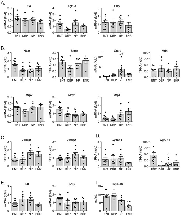
Background: Clinical reports show a positive correlation between phytosterol concentrations and severity of cholestatic liver disease markers in infants during long-term administration of parenteral lipid emulsions. Establishing a causal link between phytosterols and cholestasis has been complicated by confounding factors of lipid emulsion load, fatty acid composition, and vitamin E in many of these studies. The goal of this study is to determine whether altering the phytosterol concentration within a common soybean oil-based emulsion will alter the onset and severity of cholestasis in parenterally fed preterm piglets.
Methods: Preterm piglets were administered, for 21 days, either enteral nutrition (ENT) or parenteral nutrition (PN) prepared from a soybean oil-based emulsion containing either 24.0% (depleted [DEP]), 100% (Intralipid; normal phytosterol [NP] concentration), or 144% (enriched [ENR]) total phytosterol concentration.
Results: At the end of the study, plasma and liver phytosterol concentrations were highest in the ENR group, followed by NP and then DEP and ENT. Serum direct bilirubin, serum bile acids, and γ-glutamyltransferase were higher in the ENR and NP groups compared with either DEP or ENT groups. All PN lipid groups showed evidence of mild hepatic steatosis but no change in hepatic expression of proinflammatory cytokines or Farnesoid X receptor target genes.
Conclusion: The increase in serum direct bilirubin was lower in the DEP group vs the lipid emulsions with normal or ENR phytosterols. Our results provide additional evidence that phytosterols are linked to an increase in serum markers of cholestasis in preterm PN-fed pigs.
Keywords: bile acids; bile salt export pump; parenteral nutrition; parenteral nutrition-associated cholestasis; phytosterols; soybean oil.
© 2021 American Society for Parenteral and Enteral Nutrition.
Conflict of interest statement
Conflict of interest: Fresenius Kabi, the manufacturer of soy oil-based emulsions, provided the modified lipid emulsions. In addition, they were involved in the design of the study and the editing and preparation of the manuscript. Fresenius Kabi was involved in histopathology scoring that was confirmed independently by pathologist at Baylor College of Medicine. The authors had final authority on inclusion of data and interpretation of results.
Figures


Similar articles
-
New generation lipid emulsions prevent PNALD in chronic parenterally fed preterm pigs.J Lipid Res. 2014 Mar;55(3):466-77. doi: 10.1194/jlr.M044545. Epub 2014 Jan 29. J Lipid Res. 2014. PMID: 24478031 Free PMC article.
-
Vitamin E in New-Generation Lipid Emulsions Protects Against Parenteral Nutrition-Associated Liver Disease in Parenteral Nutrition-Fed Preterm Pigs.JPEN J Parenter Enteral Nutr. 2016 Jul;40(5):656-71. doi: 10.1177/0148607114567900. Epub 2015 Jan 16. JPEN J Parenter Enteral Nutr. 2016. PMID: 25596209 Free PMC article.
-
Mixed Lipid, Fish Oil, and Soybean Oil Parenteral Lipids Impact Cholestasis, Hepatic Phytosterol, and Lipid Composition.J Pediatr Gastroenterol Nutr. 2019 Jun;68(6):861-867. doi: 10.1097/MPG.0000000000002313. J Pediatr Gastroenterol Nutr. 2019. PMID: 30889135
-
Lipid emulsions for parenterally fed preterm infants.Cochrane Database Syst Rev. 2019 Jun 4;6(6):CD013163. doi: 10.1002/14651858.CD013163.pub2. Cochrane Database Syst Rev. 2019. PMID: 31158919 Free PMC article.
-
Impact of Parenteral Lipid Emulsion Components on Cholestatic Liver Disease in Neonates.Nutrients. 2021 Feb 4;13(2):508. doi: 10.3390/nu13020508. Nutrients. 2021. PMID: 33557154 Free PMC article. Review.
Cited by
-
Depletion of phytosterols from intravenous lipid emulsions: to be or not to be.Pediatr Res. 2025 Jun;97(7):2179-2181. doi: 10.1038/s41390-025-03877-6. Epub 2025 Feb 3. Pediatr Res. 2025. PMID: 39900833 Free PMC article.
-
Expression of circadian regulatory genes is dysregulated by increased cytokine production in mice subjected to concomitant intestinal injury and parenteral nutrition.PLoS One. 2023 Aug 30;18(8):e0290385. doi: 10.1371/journal.pone.0290385. eCollection 2023. PLoS One. 2023. PMID: 37647292 Free PMC article.
-
Selective Agonism of Liver and Gut FXR Prevents Cholestasis and Intestinal Atrophy in Parenterally Fed Neonatal Pigs.bioRxiv [Preprint]. 2024 Sep 7:2024.09.03.611073. doi: 10.1101/2024.09.03.611073. bioRxiv. 2024. PMID: 39282416 Free PMC article. Preprint.
-
A Medium-Chain Fatty Acid Analogue Prevents Intestinal Failure-Associated Liver Disease in Preterm Yorkshire Piglets.Gastroenterology. 2023 Sep;165(3):733-745.e9. doi: 10.1053/j.gastro.2023.05.035. Epub 2023 May 30. Gastroenterology. 2023. PMID: 37263310 Free PMC article.
References
-
- Carter BA, Shulman RJ. Mechanisms of disease: update on the molecular etiology and fundamentals of parenteral nutrition associated cholestasis. Nat.Clin.Pract.Gastroenterol.Hepatol 2007;4(5):277–287. - PubMed
-
- Christensen RD, Henry E, Wiedmeier SE, Burnett J, Lambert DK. Identifying patients, on the first day of life, at high-risk of developing parenteral nutrition-associated liver disease. J Perinatol. May 2007;27(5):284–290. - PubMed
-
- Ostlund RE Jr. Phytosterols in human nutrition. Annu Rev Nutr. 2002;22:533–549. - PubMed
-
- Gura KM, Duggan CP, Collier SB, et al. Reversal of parenteral nutrition-associated liver disease in two infants with short bowel syndrome using parenteral fish oil: implications for future management. Pediatrics. Jul 2006;118(1):e197–201. - PubMed
Publication types
MeSH terms
Substances
Grants and funding
LinkOut - more resources
Full Text Sources
Other Literature Sources
Research Materials
Miscellaneous
