Review
. 2021 Jul;81(5):568-590.
doi: 10.1002/dneu.22814. Epub 2021 Mar 8.

Microglia regulate synaptic development and plasticity

Affiliations
Review

Microglia regulate synaptic development and plasticity

Megumi Andoh et al. Dev Neurobiol. 2021 Jul.

Abstract

Synapses are fundamental structures of neural circuits that transmit information between neurons. Thus, the process of neural circuit formation via proper synaptic connections shapes the basis of brain functions and animal behavior. Synapses continuously undergo repeated formation and elimination throughout the lifetime of an organism, reflecting the dynamics of neural circuit function. The structural transformation of synapses has been described mainly in relation to neural activity-dependent strengthening and weakening of synaptic functions, that is, functional plasticity of synapses. An increasing number of studies have unveiled the roles of microglia, brain-resident immune cells that survey the brain parenchyma with highly motile processes, in synapse formation and elimination as well as in regulating synaptic function. Over the past 15 years, the molecular mechanisms underlying microglia-dependent regulation of synaptic plasticity have been thoroughly studied, and researchers have reported that the disruption of microglia-dependent regulation causes synaptic dysfunction that leads to brain diseases. In this review, we will broadly introduce studies that report the roles of microglia in synaptic plasticity and the possible underlying molecular mechanisms.

Keywords: microglia; synapse competition; synapse elimination; synapse engulfment; synapse formation.


Conflict of interest statement

The authors declare no conflicts of interests.

Figures
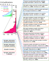
FIGURE 1
FIGURE 1
Groundbreaking studies on the microglia–synapse interaction. Some of the essential groundbreaking studies in the history of microglial research are listed in chronological order. These studies help us deeply discuss the mechanisms underlying the regulation of synaptic development and plasticity by microglia. These works improved our comprehension of microglia–synapse interactions, which is reflected by an explosive increase in related papers over the past 15 years. The number of publications was analyzed using PubMed
FIGURE 2
FIGURE 2
Visualization of the microglia–synapse interaction. (a) Iba1‐immunostained microglia and a GFP‐labeled neuron in the mouse dentate gyrus. (b) A representative image of the microglia–spine interaction. (c) A single plane of the magnified image in b. (d) Illustration of players in synaptic phagocytosis by microglia. It should be noted that the environment surrounding microglial‐synaptic phagocytosis consists of not only microglia and spines but also presynaptic boutons, astrocytes, and extracellular matrix as well as several molecules related to synaptic phagocytosis
FIGURE 3
FIGURE 3
Synapse elimination pathways that are excessively enhanced in neurological disorders. Some neurological disorders induce complement expression and microglial activation, leading to synaptic phagocytosis and spine loss by microglia
FIGURE 4
FIGURE 4
Possible molecular mechanisms by which microglia determine which synapse to phagocytose. Extracellular matrix: Neuronal activity modulates the formation and degradation of extracellular matrix, which affects the ability of microglia to approach synapses. Astrocytes: When neuronal activity is elevated, the area of astrocytes covering synapses is increased, which affects the ability of microglia to approach synapses. Norepinephrine: Norepinephrine is released in an activity‐dependent manner, inducing retraction of microglial processes from synapses. ICAM‐5: ICAM‐5 is released in an activity‐dependent manner, suppressing microglial adhesion to surrounding tissues and phagocytic activity. ROS: ATP demand decreases in synapses with low activity, which promotes ROS production, leading to the expression of PS on synapse surfaces

References

    1. Adaikkan, C., Middleton, S. J., Marco, A., Pao, P.‐C., Mathys, H., Kim, D.‐W., Gao, F., Young, J. Z., Suk, H.‐J., Boyden, E. S., McHugh, T. J., & Tsai, L.‐H. (2019). Gamma entrainment binds higher‐order brain regions and offers neuroprotection. Neuron, 102, 929–943.e928. 10.1016/j.neuron.2019.04.011 - DOI - PMC - PubMed
    1. Andoh, M., Ikegaya, Y., & Koyama, R. (2019. a). Microglia as possible therapeutic targets for autism spectrum disorders. Progress in Molecular Biology and Translational Science, 167, 223–245. 10.1016/bs.pmbts.2019.06.012 - DOI - PubMed
    1. Andoh, M., Ikegaya, Y., & Koyama, R. (2019. b). Synaptic pruning by microglia in epilepsy. Journal of Clinical Medicine, 8, 2170. 10.3390/jcm8122170. - DOI - PMC - PubMed
    1. Andoh, M., Ikegaya, Y., & Koyama, R. (2020). Microglia in animal models of autism spectrum disorders. Progress in Molecular Biology and Translational Science, 173, 239–273. 10.1016/bs.pmbts.2020.04.012 - DOI - PubMed
    1. Andoh, M., Shibata, K., Okamoto, K., Onodera, J., Morishita, K., Miura, Y., Ikegaya, Y., & Koyama, R. (2019). Exercise reverses behavioral and synaptic abnormalities after maternal inflammation. Cell Reports, 27, 2817–2825.e2815. 10.1016/j.celrep.2019.05.015 - DOI - PubMed

Publication types