

Cone Beam Computed Tomography Analysis of Sphenoid Sinus Pneumatization and Relationship with Neurovascular Structures
- PMID: 33584051
- PMCID: PMC7855127
- DOI: 10.1007/s12663-020-01326-x
Cone Beam Computed Tomography Analysis of Sphenoid Sinus Pneumatization and Relationship with Neurovascular Structures
Abstract
Background: The sphenoid sinus is considered as the most variable pneumatized structure of the skull.
Purpose: The aim of the present study was to determine the prevalence of the Onodi cell as well as to evaluate the relationship between the sphenoid sinus type of pneumatization and the presence of surrounding neurovascular protrusion using cone beam computed tomography (CBCT).
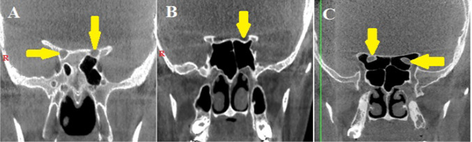
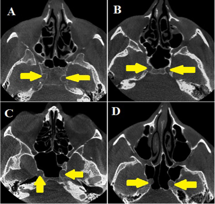
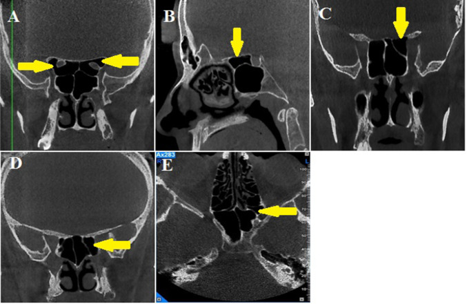
Methods: The CBCT images of 500 patients/996 sides [203 males (40.6%) and 297 females (59.4%)] were analyzed in this study. The type of sphenoid sinus pneumatization, prevalence of internal carotid artery (ICA) and optic nerve (ON) protrusion and dehiscence, and also the frequency of Onodi cell were assessed.
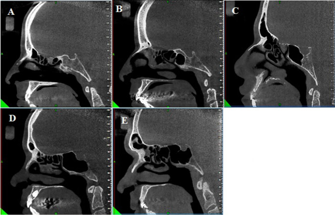
Results: The percentages of the conchal, presellar, sellar, postsellar (a), and postsellar (b) types of pneumatization were 1%, 11.5%, 35.5%, 38.9%, and 13.1%, respectively. The more the sphenoid sinuses pneumatized, the greater the frequency of ON and ICA protrusion and dehiscence of their wall to the sinus. The prevalence of Onodi cell was 38.8%. A significant correlation was found between ON dehiscence and the presence of Onodi cells.
Conclusion: The present study demonstrated a significant relationship between the sinus type and frequency of neurovascular protrusions. Therefore, the sphenoid sinus extent of pneumatization might be useful in predicting the risk of iatrogenic damage to the surrounding structures.
Keywords: Cone beam computed tomography; Internal carotid artery; Optic nerve; Pneumatization; Sphenoid sinus.
© The Association of Oral and Maxillofacial Surgeons of India 2020.
Conflict of interest statement
Conflict of interestAll authors declare that they have no conflict of interest.
Figures
Similar articles
-
Sphenoid Sinus Pneumatization Types and Correlation with Adjacent Neurovascular Structures Using Cone-Beam Computed Tomography.Indian J Otolaryngol Head Neck Surg. 2023 Sep;75(3):2245-2250. doi: 10.1007/s12070-023-03796-0. Epub 2023 May 10. Indian J Otolaryngol Head Neck Surg. 2023. PMID: 37636739 Free PMC article.
-
Sphenoid sinus types, dimensions and relationship with surrounding structures.Ann Anat. 2016 Jan;203:69-76. doi: 10.1016/j.aanat.2015.02.013. Epub 2015 Mar 20. Ann Anat. 2016. PMID: 25843780
-
A Comprehensive Computed Tomographic Analysis of Pneumatization Pattern of Sphenoid Sinus and Their Association with Protrusion/Dehiscence of Vital Neurovascular Structure in a Pakistani Subgroup.Turk Neurosurg. 2023;33(3):501-508. doi: 10.5137/1019-5149.JTN.40154-22.3. Turk Neurosurg. 2023. PMID: 36951035
-
Identification of Onodi cell and new classification of sphenoid sinus for endoscopic sinus surgery.Int Forum Allergy Rhinol. 2015 Nov;5(11):1068-76. doi: 10.1002/alr.21567. Epub 2015 Jun 10. Int Forum Allergy Rhinol. 2015. PMID: 26097234 Review.
-
Anatomical variations of the sphenoid sinus and its adjacent structures: a review of existing literature.Surg Radiol Anat. 2014 Jul;36(5):419-27. doi: 10.1007/s00276-013-1214-1. Epub 2013 Oct 22. Surg Radiol Anat. 2014. PMID: 24146215 Review.
Cited by
-
Evaluation of the relationship between sphenoid sinus morphology and area and volume by computed tomography.Oral Radiol. 2024 Apr;40(2):138-147. doi: 10.1007/s11282-023-00711-9. Epub 2023 Sep 25. Oral Radiol. 2024. PMID: 37749336
-
Computed tomography based evaluation of the association between sphenoid sinus pneumatization patterns and variations of adjacent bony structures in relation to age and gender.Neurosurg Rev. 2024 Jul 24;47(1):349. doi: 10.1007/s10143-024-02594-8. Neurosurg Rev. 2024. PMID: 39046640 Free PMC article.
-
Correlation between sphenoid sinus pneumatization and sella turcica dimensions using computed tomography.J Int Med Res. 2024 Oct;52(10):3000605241287021. doi: 10.1177/03000605241287021. J Int Med Res. 2024. PMID: 39435554 Free PMC article.
-
Sphenoid Sinus Pneumatization Types and Correlation with Adjacent Neurovascular Structures Using Cone-Beam Computed Tomography.Indian J Otolaryngol Head Neck Surg. 2023 Sep;75(3):2245-2250. doi: 10.1007/s12070-023-03796-0. Epub 2023 May 10. Indian J Otolaryngol Head Neck Surg. 2023. PMID: 37636739 Free PMC article.
-
Surgical-related Morphological Characteristics of Sphenoid Sinuses: A Comprehensive CT-Based Analysis.J Clin Exp Dent. 2024 Dec 1;16(12):e1445-e1453. doi: 10.4317/jced.62099. eCollection 2024 Dec. J Clin Exp Dent. 2024. PMID: 39822784 Free PMC article.
References
-
- Stankiewicz JA. Complications of endoscopic nasal surgery: occurrence and treatment. Am J Rhinol. 1987;1:45–49. doi: 10.2500/105065887781390417. - DOI
LinkOut - more resources
Full Text Sources
Miscellaneous