. 2021 Jan 27:11:592238.
doi: 10.3389/fphar.2020.592238. eCollection 2020.

Nasal Delivery of Hesperidin/Chitosan Nanoparticles Suppresses Cytokine Storm Syndrome in a Mouse Model of Acute Lung Injury

Affiliations

Nasal Delivery of Hesperidin/Chitosan Nanoparticles Suppresses Cytokine Storm Syndrome in a Mouse Model of Acute Lung Injury

Hua Jin et al. Front Pharmacol. .

Abstract

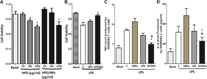
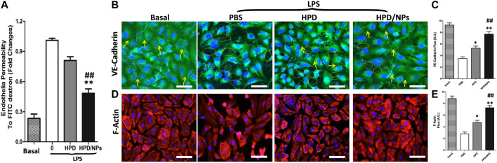
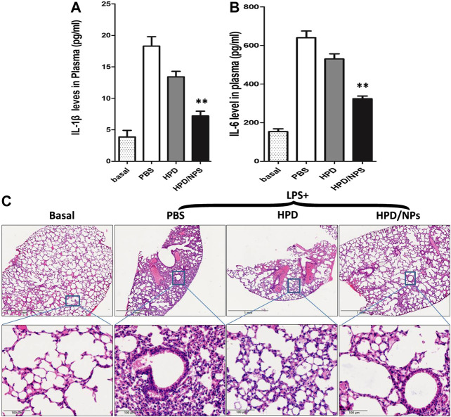
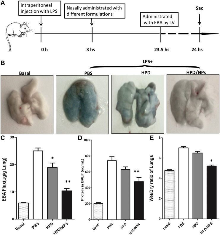
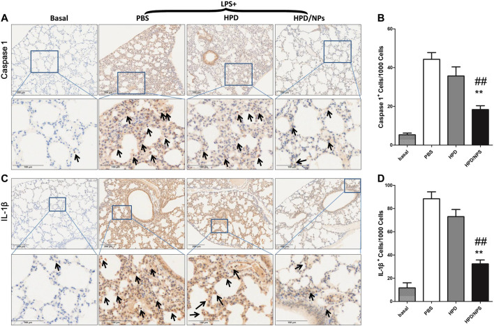
The cytokine storm or cytokine storm syndrome (CSS) is associated with high mortality in patients with acute lung injury (ALI) and acute respiratory distress syndrome (ARDS), for example following sepsis or infectious diseases including COVID-19. However, there are no effective treatments for CSS-associated ALI or ALI/ARDS. Thus, there remains an urgent need to develop effective drugs and therapeutic strategies against CSS and ALI/ARDS. Nasal and inhaled drug delivery methods represent a promising strategy in the treatment of inflammatory lung disease as a result of their ability to improve drug delivery to lungs. Improving the nasal mucosa absorption of poorly water-soluble drugs with poor mucosa bioavailability to a therapeutically effective level is another promising strategy in the fight against ALI/ARDS. Here, chitosan nanoparticles loaded with hesperidin (HPD/NPs) were developed for nasal delivery of the anti-inflammatory HPD compound to inflammatory lungs. In vitro and in vivo, HPD/NPs exhibited enhanced cellular uptake in the inflammatory microenvironment compared with free HPD. In a mouse model of inflammatory lung disease, the HPD/NPs markedly inhibited lung injury as evidenced by reduced inflammatory cytokine levels and suppressed vascular permeability compared with free HPD. Collectively, our study demonstrates that nasal delivery of HPD/NPs suppresses CSS and ALI/ARDS in a murine model of inflammatory lung disease, and that nanoparticle-based treatment strategies with anti-inflammatory effects could be used to reduce CSS and ALI in patients with inflammatory lung injury.

Keywords: chitosan nanoparticle; cytokine storm syndrome; hesperidin; lung inflammation; nasal drug delivery.


Conflict of interest statement

The authors declare that the research was conducted in the absence of any commercial or financial relationships that could be construed as a potential conflict of interest.

Figures

FIGURE 1
FIGURE 1
Characterization of HPD/NPs. (A) Scanning electron microscopy (SEM) image of an HPD/NP in suspension. (B) Mean size and (C) zeta potentials of HPD/NPs. (D) HPD/NP size over time (n = 4/group). (E) In vitro release of HPD from HPD/NPs in PBS (0.01 M, PH = 7.4, n = 4/group).
FIGURE 2
FIGURE 2
Impact of HPD and HPD/NPs on LPS-induced inflammatory response in vitro. (A). Viability of RAW264.7 macrophages exposed to different doses of HPD or HPD/NPs for 24 h. (B) Viability of RAW264.7 macrophages exposed to 10 mg/ml HPD or HPD/NPs or vehicle (0.02% DMSO) for 3 h, followed by stimulation with 1 µg/ml of LPS for 24 h. (C) NO and (D) IL-6 production by RAW264.7 macrophages exposed to 10 mg/ml HPD or HPD/NPs or vehicle (0.02% DMSO) for 3 h, followed by stimulation with 1 µg/ml of LPS for 24 h (A) N = 4/group, *p < 0.05 vs. basal group or 10 μg/ml group. (B–D). N = 4/group, *p < 0.05 vs. vehicle control; #p < 0.05 vs. HPD group.
FIGURE 3
FIGURE 3
Impact of HPD and HPD/NPs on LPS-induced endothelial integrity in vitro. (A) Permeability of the HUVEC monolayer to FITC-dextran (40 kDa) after 3 h exposure to PBS or HPD or HPD/NPs (10 µg/ml) followed by 24 h LPS exposure. Data expressed as a percentage of PBS + LPS treatment group (n = 4/group, **p < 0.001 vs. vehicle control group; ##p < 0.001 vs. HPD group). (B) Representative images and (C) quantification of immunostaining for VE-cadherin (green) in HUVEC monolayers Arrows indicate VE-cadherin-positive cell junctions; circles indicate weak or absent VE-cadherin signal at cell junction. (D) Representative images and (E) quantification of F-actin (red) immunostaining of HUVECs after 3 h exposure to PBS or HPD or HPD/NPs followed by 24 h LPS (3.5 mg/kg). Nuclei were counterstained with DAPI (blue). Scale bar, 50 μm *p < 0.05 and **p < 0.01 vs. PBS group; ##p < 0.01 vs. HPD group. A.U., arbitrary units.
FIGURE 4
FIGURE 4
Impact of HPD and HPD/NPs on LPS-induced inflammation in mice. At 3 h postLPS, PBS (vehicle), HPD, or HPD/NPs were nasally administered to mice. Lung tissues were collected at 24 h post-LPS challenge. (A) Expression levels of IL-1β and (B) IL-6 in mouse plasma at 24 h post-LPS challenge (n = 4/group; **p < 0.001 vs. PBS vehicle and vs. HPD). (C) Representative micrographs of H&E stained lung tissue cross-sections at 24 h post-LPS challenge. Scale bar, 1 mm (upper row) or 100 μm (lower row).
FIGURE 5
FIGURE 5
Impact of HPD and HPD/NPs on LPS-induced ALI in mice. At 3 h post-LPS, PBS (vehicle), HPD, or HPD/NPs were nasally administered to mice. Lung tissues were collected at 24 h post-LPS challenge. (A) EBA assay schematic. (B) Representative images of murine lung tissues after EBA-injection. (C) EBA flux, (D) BAL protein, and (E) wet/dry weight ratio. (C–E) N = 5; *p < 0.05 and **p < 0.001 vs. PBS group.
FIGURE 6
FIGURE 6
Impact of HPD and HPD/NPs on markers of pyroptosis in lungs of LPS-challenged mice. At 3 h post-LPS, PBS (vehicle), HPD, or HPD/NPs were nasally administered to mice. Lung tissues were collected at 24 h post-LPS challenge. (A) Representative images and (B) quantification of lung tissue cross-sections immune-stained for caspase 1. (C) Representative images and (D) quantification of lung tissue cross-sections immune-stained for IL-1β. Arrows indicate brown/positive staining. N = 4/group; **p < 0.01 vs. PBS, ## p < 0.01 vs. HPD.

References

    1. Bergsbaken T., Fink S. L., Cookson B. T. (2009). Pyroptosis: host cell death and inflammation. Nat. Rev. Microbiol., 7 (2), 99–109. 10.1038/nrmicro2070 - DOI - PMC - PubMed
    1. Bruinsmann F. A., Pigana S., Aguirre T., Dadalt Souto G., Garrastazu Pereira G., Bianchera A., et al. (2019). Chitosan-coated nanoparticles: effect of chitosan molecular weight on nasal transmucosal delivery. Pharmaceutics 11 (2), 86 10.3390/pharmaceutics11020086 - DOI - PMC - PubMed
    1. Channappanavar R., Perlman S. (2017). Pathogenic human coronavirus infections: causes and consequences of cytokine storm and immunopathology. Semin. Immunopathol., 39 (5), 529–539. 10.1007/s00281-017-0629-x - DOI - PMC - PubMed
    1. Ciftci O., Ozcan C., Kamisli O., Cetin A., Basak N., Aytac B. (2015). Hesperidin, a citrus flavonoid, has the ameliorative effects against experimental autoimmune encephalomyelitis (EAE) in a C57BL/J6 mouse model. Neurochem. Res., 40 (6), 1111–1120. 10.1007/s11064-015-1571-8 - DOI - PubMed
    1. Du Z. A., Sun M. N., Hu Z. S. (2018). Saikosaponin a ameliorates LPS-induced acute lung injury in mice. Inflammation, 41 (1), 193–198. 10.1007/s10753-017-0677-3 - DOI - PubMed

LinkOut - more resources