. 2021 Jan 27:11:614450.
doi: 10.3389/fphar.2020.614450. eCollection 2020.

Cynandione A Alleviates Neuropathic Pain Through α7-nAChR-Dependent IL-10/β-Endorphin Signaling Complexes

Affiliations

Cynandione A Alleviates Neuropathic Pain Through α7-nAChR-Dependent IL-10/β-Endorphin Signaling Complexes

Qiao-Qiao Han et al. Front Pharmacol. .

Abstract

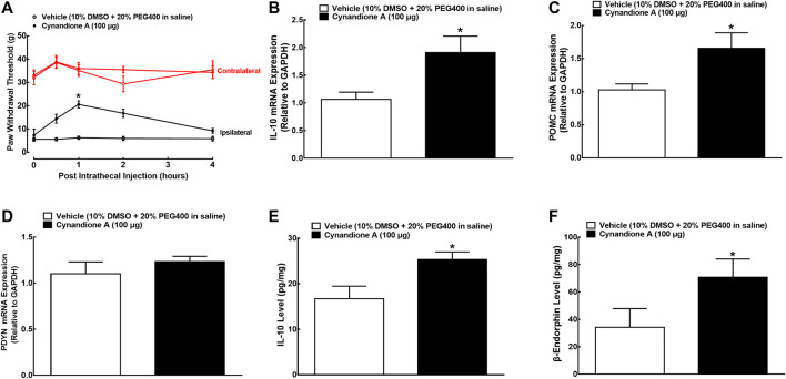
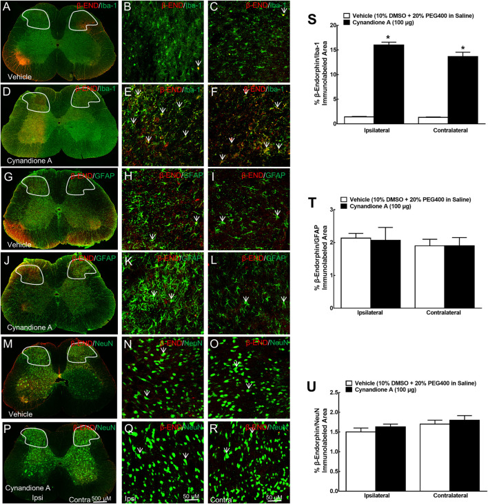
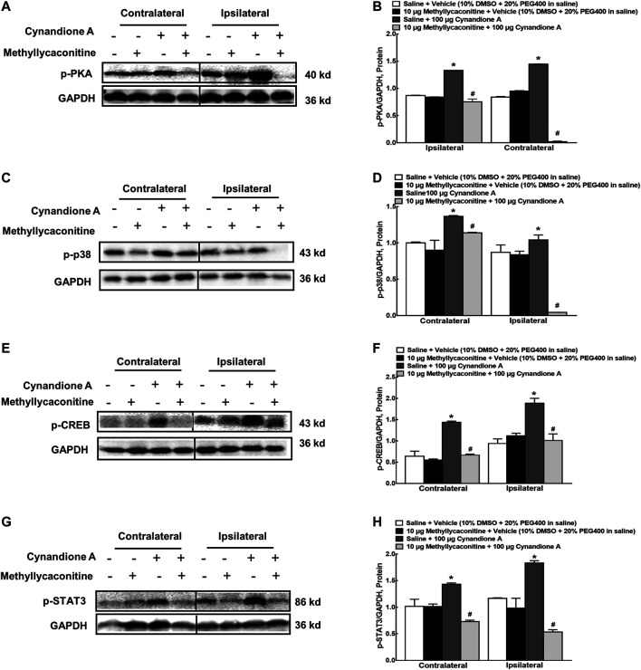
Cynandione A, an acetophenone isolated from Cynanchum Wilfordii Radix, exhibits antineuropathic pain effect. This study further explored the target molecule and signaling mechanisms underlying cynandione-A-induced antineuropathic pain. Intrathecal injection of cynandione A significantly attenuated mechanical allodynia in neuropathic rats and substantially increased spinal expression of IL-10 and β-endorphin but not dynorphin A. Cynandione A treatment also enhanced expression of IL-10 and β-endorphin but not α7 nicotinic acetylcholine receptors (nAChRs) in cultured microglia. The IL-10 antibody attenuated cynandione-A-induced spinal or microglial gene expression of β-endorphin and mechanical allodynia, whereas the β-endorphin antiserum blocked cynandione-A-induced mechanical antiallodynia but not spinal or microglial IL-10 gene expression. The α7 nAChR antagonist methyllycaconitine significantly reduced cynandione-A-induced mechanical antiallodynia and spinal or microglial expression of IL-10 and β-endorphin. Furthermore, cynandione A stimulated microglial phosphorylation of PKA, p38, and CREB in an α7-nAChR-dependent manner, and treatment with their inhibitors attenuated cynandione-A-induced mechanical antiallodynia and spinal or microglial expression of IL-10 and β-endorphin. In addition, cynandione A stimulated spinal phosphorylation of the transcription factor STAT3, which was inhibited by methyllycaconitine, the PKA activation inhibitor or IL-10 antibody. The STAT3 inhibitor NSC74859 also abolished cynandione-A-induced mechanical antiallodynia and spinal expression of β-endorphin. These findings suggest that cynandione A suppresses neuropathic pain through α7-nAChR-dependent IL-10/β-endorphin signaling pathway in spinal microglia.

Keywords: cynandione A; interleukin-10; microglia; neuropathic pain; α7 nicotinic acetylcholine receptor; β-endorphin.


Conflict of interest statement

The authors declare that the research was conducted in the absence of any commercial or financial relationships that could be construed as a potential conflict of interest.

Figures

FIGURE 1
FIGURE 1
Effects of cynandione A, given intrathecally, on mechanical allodynia (A), spinal mRNA expression of IL-10 (B), the β-endorphin precursor proopiomelanocortin (POMC (C)) and dynorphin precursor prodynorphin (PDYN (D)), and spinal protein expression of IL-10 (E) and β-endorphin (F) in neuropathic rats induced by L5/L6 spinal nerve ligation. Spinal lumbar enlargements (L3-L5) were obtained from the sacrificed rats 1 h after injection of cynandione A or the vehicle. The gene and protein expression was determined by using qRT-PCR and enzyme-linked immunosorbent fluorescent assays, respectively. Data are shown as means ± SEM (n = 6 per group). * p < 0.05 compared with the vehicle group, analyzed by two-tailed and unpaired Student t-test or repeated measures two-way ANOVA followed by the post-hoc Student-Newman-Keuls test.
FIGURE 2
FIGURE 2
Specific stimulatory effects of cynandione A, given intrathecally, on spinal microglial expression of IL-10 in neuropathic rats induced by tight ligation of L5/L6 spinal nerves. Neuropathic rats that received intrathecal injection of cynandione A or the vehicle were sacrificed 1 h after injection and the spinal lumbar enlargements (L3–L5) were obtained. The specific expression of IL-10 was double fluorescence immunolabeled with the microglial cellular biomarker Iba-1 (A–F), astrocytic cellular biomarker GFAP (G-L), and neuronal cellular biomarker NeuN (M–R) in the contralateral and ipsilateral spinal cord and dorsal horn I-Ⅲ laminate. Arrows indicate yellow double labeling of IL-10 with the corresponding cellular biomarkers. The immunolabeled surface areas of IL-10/Iba-1 (S), IL-10/GFAP (T), and IL-10/NeuN (U) from the indicated spinal dorsal horn laminae I-Ⅲ were quantified using the Image J program. Data are presented as means ± SEM (n = 5 per group). * P < 0.05 compared with the vehicle group, analyzed by one-way ANOVA followed by the post-hoc Student-Newman-Keuls test.
FIGURE 3
FIGURE 3
Specific stimulatory effects of cynandione A, given intrathecally, on spinal microglial expression of β-endorphin in neuropathic rats induced by tight ligation of L5/L6 spinal nerves. Neuropathic rats that received intrathecal injection of cynandione A or the vehicle were sacrificed 1 h after injection and the spinal lumbar enlargements (L3–L5) were obtained. Expression of β-endorphin was double fluorescence immunolabeled with the microglial cellular biomarker Iba-1 (A–F), astrocytic cellular biomarker GFAP (G-L), and neuronal cellular biomarker NeuN (M–R) in the contralateral and ipsilateral spinal cord and dorsal horn I-Ⅲ laminate. Arrows indicate yellow double labeling of β-endorphin with the corresponding cellular biomarkers. The immunolabeled surface areas of β-endorphin/Iba-1 (S), β-endorphin/GFAP (T), and β-endorphin/NeuN (U) from the indicated spinal dorsal horn laminae I-Ⅲ were quantified using the Image J program. Data are presented as means ± SEM (n = 5 per group). * P < 0.05 compared with the vehicle group, analyzed by one-way ANOVA followed by the post-hoc Student-Newman-Keuls test.
FIGURE 4
FIGURE 4
Effects of cynandione A on gene (A–C) expression, protein secretion (D, E), and fluorescent immunostaining (F–S) of IL-10, β-endorphin, and dynorphin A in primary cultures of spinal microglia collected from 1-day-old neonatal rats. Primary microglial cells were incubated with cynandione A for 2 h. The qRT-PCR and enzyme-linked immunosorbent fluorescent kits were used to detect gene expression and protein secretion in the cells and culture medium, respectively. Single and triple immunofluorescence staining was used to detect I-10 (red), Iba-1 (green), and IL-10 (red)/Iba-1 (green)/DAPI (blue) or β-endorphin (red)/Iba-1 (green)/DAPI (blue). The immunolabeled surface areas of IL-10/Iba-1 (R) and β-endorphin/Iba-1 (S) were quantified by using the Image J program. The data are presented as means ± SEM (n = 3 per group with two independent repeats). * p < 0.05 compared with the vehicle group, by two-tailed and unpaired Student t-test.
FIGURE 5
FIGURE 5
Blockade effects of the IL-10 antibody and β-endorphin antiserum on cynandione-A-induced spinal mechanical antiallodynia (A), spinal gene expression of IL-10 (B) and the β-endorphin precursor proopiomelanocortin (POMC (C)), and spinal protein expression of IL-10 (D) and β-endorphin (E) in neuropathic rats induced by L5/L6 spinal nerve ligation. Neuropathic rats received intrathecal injection of saline, the IL-10 antibody or β-endorphin antiserum followed by intrathecal injection of the vehicle or cynandione A 30 min later. The spinal cords were obtained immediately after the completion of behavioral test (1 h after the last injection) for detection of IL-10 and β-endorphin by using qRT-PCR and enzyme-linked immunosorbent fluorescent assays, respectively. Blockade effects of the IL-10 antibody and β-endorphin antiserum on cynandione-A-stimulated gene expression of POMC (F) and IL-10 (G) in cultured microglial cells originated from 1-day-old neonatal rats. Data are shown as means ± SEM (n = 6 per group in neuropathic rats or n = 3 per group with two independent repeats in cultured cells). *, # p < 0.05 compared with the control and cynandione A treatment groups, respectively, analyzed by one-way ANOVA followed by the post-hoc Student-Newman-Keuls test.
FIGURE 6
FIGURE 6
Blockade effects of the α7 nicotinic acetylcholine receptor (α7 nAChR) antagonist methyllycaconitine, given before or after cynandione A injection, on cynandione-A-induced spinal mechanical antiallodynia in the ipsilateral hindpaws (A,B), spinal gene expression of IL-10 (C) and the β-endorphin precursor proopiomelanocortin (POMC (D)), and protein expression of IL-10 (E) and β-endorphin (F) in neuropathic rats induced by spinal nerve ligation. Neuropathic rats received intrathecal injection of the vehicle or methyllycaconitine followed by intrathecal saline or cynandione A 30 min later. The spinal cords were obtained 1 h after the last intrathecal injection for detection of IL-10 and β-endorphin by using qRT-PCR and enzyme-linked immunosorbent fluorescent assays, respectively. For the postinjection study, neuropathic rats received intrathecal injection of cynandione A followed by methyllycaconitine 30 min later. Data are shown as means ± SEM (n = 6 per group in neuropathic rats). *, # p < 0.05 compared with the control and cynandione A treatment groups, respectively, analyzed by one-way or measures-repeated two-way ANOVA followed by the post-hoc Student-Newman-Keuls test.
FIGURE 7
FIGURE 7
Stimulatory effects of cynandione A on expression of IL-10 colocalized with the α7 nicotinic acetylcholine receptor (α7 nAChR) (A–I) and blockade effects of α7 nAChR antagonist methyllycaconitine on cynandione A-stimulated expression of IL-10 and β-endorphin (J–M) in cultured microglial cells originated from 1-day-old neonatal rats. Cultured primary microglial cells were collected 2 h after cynandione A incubation. The single and triple immunofluorescence staining of IL-10 (red), α7 nAChR (green), and IL-10 (red)/α7 nAChR (green)/DAPI (blue) was performed. The immunolabeled surface areas of IL-10 (G), α7 nAChR (H), and IL-10/α7 nAChR/DAPI (I) were quantified by using the Image J program. The qRT-PCR and enzyme-linked immunosorbent fluorescent kits were used to detect gene expression and protein secretion in the cells and culture medium, respectively. Data are presented as means ± SEM (n = 3 per group with two independent repeats). * P < 0.05 compared with the control group, analyzed by two-tailed and unpaired Student t-test or one-way followed by the post-hoc Student-Newman-Keuls test.
FIGURE 8
FIGURE 8
Blockade effects of the adenylyl cyclase inhibitor DDA, PKA activation inhibitor H-89, p38 activation inhibitor SB203580, and CREB activation inhibitor KG501 on cynandione-A-induced spinal mechanical antiallodynia and mRNA expression of IL-10 and the β-endorphin precursor proopiomelanocortin (POMC (C)) in the spinal cords of neuropathic rats induced by spinal nerve ligation (A–C) and cultured primary microglial cells originated from 1-day-old neonatal rats (D, E). Neuropathic rats received intrathecally the vehicle or DDA, H-89, SB203580 and KG501 30 min followed by cynandione A. The spinal cords and cultured microglial cells were obtained 1 h after the last intrathecal injection or 2 h after incubation to detect IL-10 and POMC gene expression by using qRT-PCR. Data are shown as means ± SEM (n = 6 per group in neuropathic rats or n = 3 per group with two independent repeats in cultured cells). *, # p < 0.05 compared with the control and cynandione A treatment groups, respectively, analyzed by one-way or two-way ANOVA followed by the post-hoc Student-Newman-Keuls test.
FIGURE 9
FIGURE 9
Blockade effects of the PKA activation inhibitor H-89 (A,B) and IL-10 antibody (C,D) on cynandione-A-stimulated spinal STAT3 phosphorylation in neuropathic rats induced by L5/L6 spinal nerve ligation. Blockade effects of the STAT3 activation inhibitor NSC74859 on cynandione-A-induced spinal mechanical antiallodynia (E) and gene expression of the β-endorphin precursor proopiomelanocortin (POMC) in the spinal cords of neuropathic rats (F) and cultured primary microglial cells originated from 1-day-old neonatal rats (G). Neuropathic rats received intrathecal H-89, the IL-10 antibody or NSC74859 30 min later followed by intrathecal cynandione A. The spinal cords were obtained 1 h after the last intrathecal injection for detection of STAT3 phosphorylation and POMC expression by using western blot and qRT-PCR, respectively. Data are shown as means ± SEM (n = 6 per group in neuropathic rats or n = 3 per group with two independent repeats in cultured cells). *, # p < 0.05 compared with the control and cynandione A treatment groups, respectively, analyzed by one-way or measures-repeated two-way ANOVA followed by the post-hoc Student-Newman-Keuls test.
FIGURE 10
FIGURE 10
Blockade effects of the α7 nicotinic acetylcholine receptor (α7 nAChR) antagonist methyllycaconitine on cynandione-A-induced phosphorylation of spinal PKA (A,B), p38 (C,D), CREB (E,F), and STAT3 (G,H) in neuropathic rats induced by L5/L6 spinal nerve ligation. Neuropathic rats received intrathecal injection of the vehicle or methyllycaconitine 30 min later followed by intrathecal cynandione A. The spinal cords were obtained 1 h after the last intrathecal injection for the phosphorylation detection by using western blot. Data are shown as means ± SEM (n = 6 per group). *, # p < 0.05 compared with the control and cynandione A treatment groups, respectively, analyzed by one-way ANOVA followed by the post-hoc Student-Newman-Keuls test.

Similar articles

Cited by

References

    1. Albuquerque E. X., Pereira E. F. R., Alkondon M., Rogers S. W. (2009). Mammalian nicotinic acetylcholine receptors: from structure to function. Physiol. Rev. 89 (1), 73–120. 10.1152/physrev.00015.2008 - DOI - PMC - PubMed
    1. Ali U., Apryani E., Wu H. Y., Mao X. F., Liu H., Wang Y. X. (2020). Low frequency electroacupuncture alleviates neuropathic pain by activation of spinal microglial IL-10/β-endorphin pathway. Biomed. Pharmacother. 125, 109898 10.1016/j.biopha.2020.109898 - DOI - PubMed
    1. Allen C. M., Ely C. M., Juaneza M. A., Parsons S. J. (1996). Activation of Fyn tyrosine kinase upon secretagogue stimulation of bovine chromaffin cells. J. Neurosci. Res. 44 (5), 421–429. 10.1002/(sici)1097-4547(19960601)44:5<421::Aid-jnr2>3.0.Co;2-h - DOI - PubMed
    1. Apryani E., Ali U., Wang Z. Y., Wu H. Y., Mao X. F., Ahmad K. A., et al. (2020). The spinal microglial IL-10/β-endorphin pathway accounts for cinobufagin-induced mechanical antiallodynia in bone cancer pain following activation of α7-nicotinic acetylcholine receptors. J. Neuroinflammation 17 (1), 75 10.1186/s12974-019-1616-z - DOI - PMC - PubMed
    1. Arredondo J., Chernyavsky A. I., Jolkovsky D. L., Pinkerton K. E., Grando S. A. (2006). Receptor-mediated tobacco toxicity: cooperation of the Ras/Raf-1/MEK1/ERK and JAK-2/STAT-3 pathways downstream of alpha7 nicotinic receptor in oral keratinocyte. Faseb. J. 20 (12), 2093–2101. 10.1096/fj.06-6191com - DOI - PubMed

LinkOut - more resources