. 2021 Jan 28:11:617149.
doi: 10.3389/fphar.2020.617149. eCollection 2020.

Triumeq Increases Excitability of Pyramidal Neurons in the Medial Prefrontal Cortex by Facilitating Voltage-Gated Ca2+ Channel Function

Affiliations

Triumeq Increases Excitability of Pyramidal Neurons in the Medial Prefrontal Cortex by Facilitating Voltage-Gated Ca2+ Channel Function

Lihua Chen et al. Front Pharmacol. .

Abstract

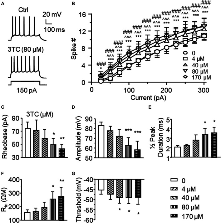
Combination antiretroviral therapy (cART) suppresses HIV-1 replication, improves immune function, and prolongs the life of people living with HIV (PLWH). However, cART also induces neurotoxicity that could complicate HIV-induced neurodegeneration while reduce its therapeutic efficacy in treating HIV/AIDS. Triumeq is a first-line cART regimen, which is co-formulated by three antiretroviral drugs (ARVs), lamivudine (3TC), abcavir (ABC), and dolutegravir (DTG). Little is known about potential side effects of ARVs on the brain (including those co-formulating Triumeq), and their mechanisms impacting neuronal activity. We assessed acute (in vitro) and chronic (in vivo) effects of Triumeq and co-formulating ARVs on pyramidal neurons in rat brain slices containing the medial prefrontal cortex (mPFC) using patch-clamp recording approaches. We found that acute Triumeq or 3TC in vitro significantly increased firing of mPFC neurons in a concentration- and time-dependent manner. This neuronal hyperactivity was associated with enhanced Ca2+ influx through voltage-gated Ca2+ channels (VGCCs). Additionally, chronic treatment with Triumeq in vivo for 4 weeks (4 wks) also significantly increased firing and Ca2+ influx via VGCCs in mPFC neurons, which was not shown after 2 wks treatment. Such mPFC neuronal hyperexcitability was not found after 4 weeks treatments of individual ARVs. Further, chronic Triumeq exposure in vivo significantly enhanced mRNA expression of low voltage-activated (LVA) L-type Ca2+ channels (Cav1.3 L-channels), while changes in high voltage-activated (HVA) Cav1.2 L-channels were not observed. Collectively, these novel findings demonstrate that chronic cART induces hyperexcitability of mPFC pyramidal neurons by abnormally promoting VGCC overactivation/overexpression of VGCCs (including, but may not limited to, LVA-Cav1.3 L-channels), which could complicate HIV-induced neurotoxicity, and ultimately may contribute to HIV-associated neurocognitive disorders (HAND) in PLWH. Determining additional target(s) of cART in mPFC pyramidal neurons may help to improve the therapeutic strategies by minimizing the side effects of cART for treating HIV/AIDS.

Keywords: CART; HIV-1; calcium dysregulation; electrophysiology; hyperactivity; neurodegenerative disease; neurotoxicity; voltage-gated calcium channel.


Conflict of interest statement

The authors declare that the research was conducted in the absence of any commercial or financial relationships that could be construed as a potential conflict of interest.

Figures

Figure 1
Figure 1
Acute treatment of lamivudine (3TC) in vitro increases firing of mPFC pyramidal neurons (A) Sample traces showing that a moderate depolarizing current pulse (150pA, which mimicked physiological excitatory inputs) evoked more action potentials in a 3TC-treated neuron compared to one without 3TC treatment (B) The current-spike relationships indicate a significant increase in the firing frequency of neurons treated with 3TC in a concentration-dependent manner (40–170 µM) compared to those without this ARV (n = 8/ea. ***p < 0.001 between 0 vs 44 µM; ^  ^^^p < 0.05 or 0.001 between 0 and 80 µM; ###p < 0.001 between 0 and 170 µM). 3TC-Associated with increased firing, higher concentrations of 3TC also altered the membrane properties by reducing the rheobase (C) and peak amplitude (D), increasing the ½ peak duration (E) and inward resistance Rin (F), while decreasing the threshold (G) (n = 8/ea. *,**,***p < 0.05, 0.01 or 0.001).
Figure 2
Figure 2
3TC enhances Ca2+ influx through overactive VGCCs (A) Sample traces showing that acute exposure to 3TC in vitro induced a prolongation of Ca2+ spikes in mPFC neurons (pointed by the arrows), indicating an increased functional activity of VGCCs in mPFC neurons (B) The acute effect of 3TC on Ca2+ spikes was also concentration-dependent, showing that the duration of Ca2+ spike was gradually increased at lower concentrations (1–80 µM), but begun to decrease in response to a high concentration (170 µM) (n = 9/ea. *,**p < 0.05 or 0.01).
Figure 3
Figure 3
Acute Triumeq treatment in vitro also increases firing of mPFC pyramidal neurons in a dose-dependent manner (A) Sample traces showing the firing numbers evoked by a moderate depolarizing current (150 pA) in mPFC neurons with or without Triumeq in the bath (B) The current-spike relationships indicate a significant increase in mPFC neuronal firing in response to 10× or 100×, but not 1×, of acute Triumeq compared to vehicle-treated controls (n = 8/ea. Triumeq effect: F 3,21 = 5.986, p = 0.004; current effect: F 11,77 = 93.88, p < 0.001; interaction: F 33,231 = 3.697, p < 0.001. post-hoc test: *,**,***p < 0.05, 0.01 or 0.001 between 0 vs 10× Triumeq; ^,^^^p < 0.05 or 0.001 between 0 vs 100× Triumeq). 1× Triumeq (containing 0.3 μg/ml ABC, 0.3 μg/ml 3TC, and 20 ng/ml DTG) equals to the concentrations of these ARVs found in the CSF of PLWH on Triumeq. 10× and 100× Triumeq refers to 10-fold and 100-fold of which, respectively.
Figure 4
Figure 4
Acute Triumeq treatment also increases VGCC activity in mPFC pyramidal neurons in a dose-dependent manner (A) Sample traces showing the acute effects of Triumeq (1×, 10×, and 100×) on evoked Ca2+ spikes (B–C) High concentrations of Triumeq in vitro significantly increased the duration (B) (n = 8/ea. F 3,21 = 7.089, p = 0.002) and the area of Ca2+ spikes (C) (n = 8/ea. F 3,21 = 4.201, p = 0.018). *p<0.05 compared to the baseline prior to acute exposure to Triumeq.
Figure 5
Figure 5
Chronic, but not subchronic, treatment of Triumeq in vivo significantly increases firing of mPFC pyramidal neurons (A) Sample traces showing firing of mPFC neurons evoked by V m depolarization after a 2 wks subchronic vehicle (upper panel) or Triumeq (lower panel) treatment in vivo (B) The current-spike relationships indicate that there was no significant difference in the firing between mPFC neurons from 2 wks Triumeq-pretreated rats and those from 2 wks vehicle-pretreated controls (n = 8/ea. Triumeq effect: F 1,14 = 0.424, p = 0.426; current effect: F 11,154 = 187.8, p < 0.001; interaction: F 11,154 = 0.418, p = 0.947) (C) Sample traces showing the changes in firing evoked by V m depolarization in mPFC neurons after 4 wks of vehicle (upper panel) or Triumeq (lower panel) treatment in vivo (D) The current-spike relationships indicate a significant increase in firing of mPFC neurons in rats after 4 wks Triumeq pretreatment compared to those in 4 wks vehicle-pretreated rats (14/ea. Triumeq effect: F 1,26 = 12.59, p = 0.002; current effect: F 11,286 = 398.9, p < 0.001; interaction: F 11,286 = 4.851, p < 0.001; post-hoc test, *,**,***p < 0.05, 0.01 or 0.001).
Figure 6
Figure 6
Chronic Triumeq treatment in vivo for 4 wks increases Ca2+ influx via VGCCs in mPFC pyramidal neurons compared to those from vehicle-pretreated controls (A) Sample traces showing Ca2+ spikes evoked by V m depolarization (indicating Ca2+ influx via VGCCs) in mPFC neurons from a vehicle-pretreated rat compared to a Triumeq-pretreated rat (B)–(C) The bar graphs indicate that the duration (B) and area (C) of Ca2+ spikes were significantly prolonged and enlarged, respectively, after a 4 wks Triumeq pretreatment (the duration: vehicle vs. Triumeq: n = 8 vs. 11; t 17 = 2.141, p = 0.047; and the area: vehicle vs. Triumeq: n = 8 vs. 11; t 17 = 2.272, p = 0.036).
Figure 7
Figure 7
Chronic Triumeq treatment in vivo for 4 wk significantly increases the mRNA expression of Cav1.3, but not Cav1.2, L-channels in the mPFC (A) The mRNA level of the Cav1.3 L-channel (Cacna1d gene) was significantly increased in the mPFC following 4 wks treatment of Triumeq compared to chronic treatment of vehicle (n = 8 rat/ea: t 14 = 2.161, p = 0.0485) (B) There was no significant change in the mRNA level of Cav1.2 (Cacna1c gene) L-channels in the mPFC after 4 wks treatment of Triumeq (n = 8 rat/ea: t 14 = 1.249, p = 0.232).
Figure 8
Figure 8
Chronic treatment of individual 3TC, ABC, or DTG alone in vivo for 4 wks does not alter firing of mPFC pyramidal neurons (A) Samples traces showing firing in response to V m hyperpolarization in mPFC neurons following 4 wks chronic treatment of SAL (upper panel), 3TC (middle panel) or ABC (lower panel) treatment in vivo (B) The current-spike relationships indicate that there was no significant difference in firing among mPFC neurons from 3TC, ABC or SAL-pretreated rats (SAL vs. 3TC: n = 9 vs. 12. 3TC effect: F (1,19) = 0.009, p = 0.925; current effect: F (11,209) = 309.6, p<0.001; interaction: F (11,206) = 0.502, p = 0.901; SAL vs. ABC: n = 9 vs. 12. ABC effect: F (1,19) = 1.136, p = 0.300; current effect: F (11,209) = 211.4, p<0.001; interaction: F (11,206) = 2.557, p = 0.005). (C) Sample traces showing the firing in response to V m hyperpolarization in mPFC neurons following 4 wks pretreatment of 70% DMSO (the solvent for DTG, upper panel), or DTG (lower panel). (D) The current-spike relationships indicate that there was no significant difference in firing between neurons from 4 wks DTG-pretreated and 4 wks DMSO-pretreated rats (n = 14/ea. DTG effect: F (1,26) = 0.587, p = 0.450; current effect: F (11,286) = 308.1, p<0.001; interaction: F (11,286) = 1.04, p = 0.411).

Similar articles

Cited by

References

    1. Akang E. N. (2019). Combination antiretroviral therapy (cART)-induced hippocampal disorders: highlights on therapeutic potential of Naringenin and Quercetin. IBRO Rep. 6, 137–146. 10.1016/j.ibror.2019.04.002 - DOI - PMC - PubMed
    1. Akay C., Cooper M., Odeleye A., Jensen B. K., White M. G., Vassoler F., et al. (2014) Antiretroviral drugs induce oxidative stress and neuronal damage in the central nervous system. J. Neurovirol. 20:39–53. 10.1007/s13365-013-0227-1 - DOI - PMC - PubMed
    1. Akay C., Lindl K. A., Wang Y., White M. G., Isaacman-Beck J., Kolson D. L., et al. (2011). Site-specific hyperphosphorylation of pRb in HIV-induced neurotoxicity. Mol. Cell. Neurosci. 47, 154–165. 10.1016/j.mcn.2011.04.001 - DOI - PMC - PubMed
    1. Antinori A., Arendt G., Becker J. T., Brew B. J., Byrd D. A., Cherner M., et al. (2007) Updated research nosology for HIV-associated neurocognitive disorders. Neurology. 69:1789–1799. 10.1212/01.WNL.0000287431.88658.8b - DOI - PMC - PubMed
    1. Aouri M., Barcelo C., Ternon B., Cavassini M., Anagnostopoulos A., Yerly S., et al. (2016). In Vivo profiling and distribution of known and novel phase I and phase II metabolites of efavirenz in plasma, urine, and cerebrospinal fluid. Drug Metab. Dispos. 44, 151–161. 10.1124/dmd.115.065839 - DOI - PubMed

LinkOut - more resources