. 2021 Jan 29:11:632809.
doi: 10.3389/fphar.2020.632809. eCollection 2020.

Loss of FGFR3 Delays Acute Myeloid Leukemogenesis by Programming Weakly Pathogenic CD117-Positive Leukemia Stem-Like Cells

Affiliations

Loss of FGFR3 Delays Acute Myeloid Leukemogenesis by Programming Weakly Pathogenic CD117-Positive Leukemia Stem-Like Cells

Chen Guo et al. Front Pharmacol. .

Abstract

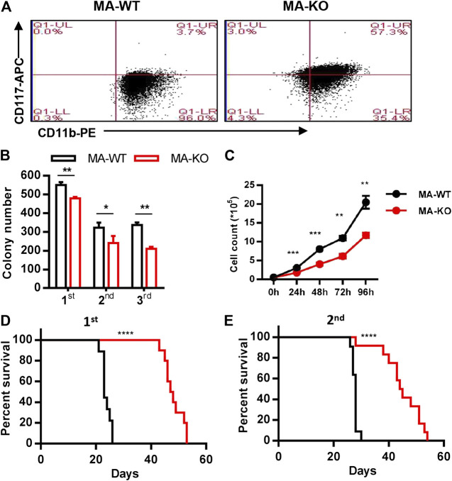
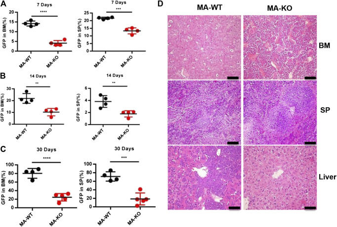
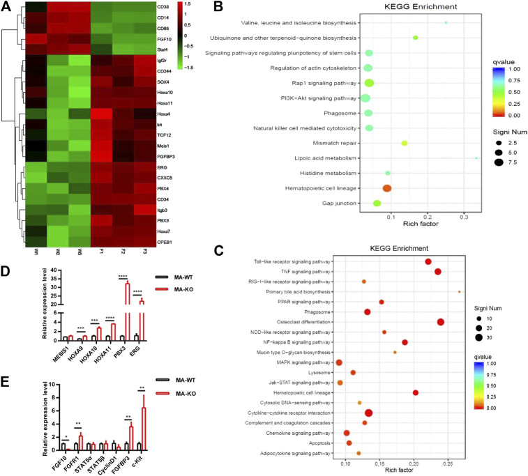
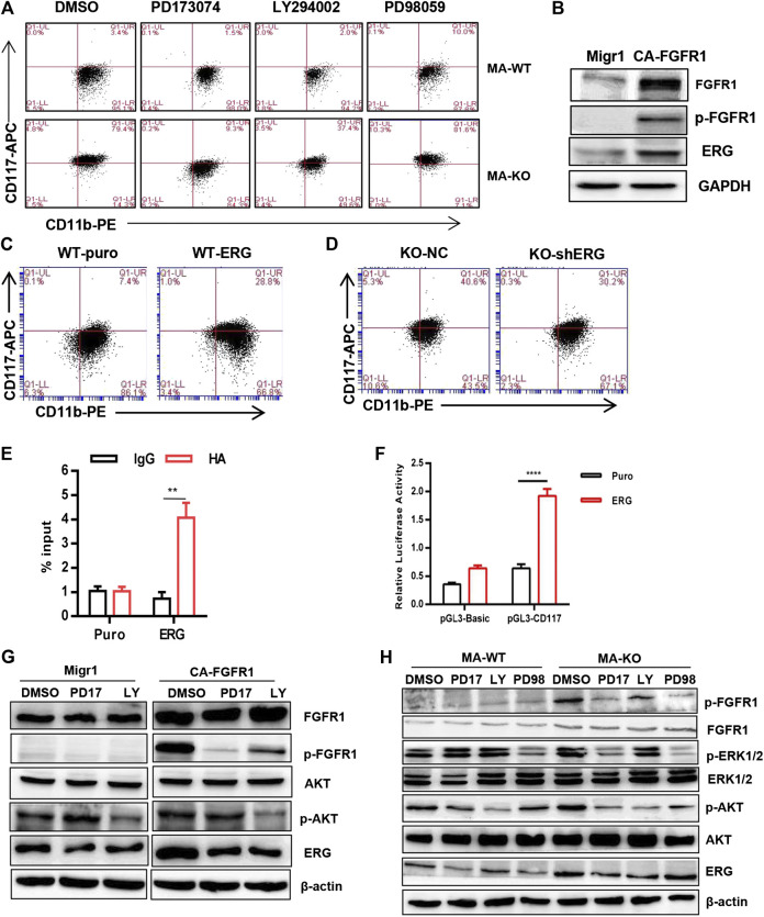
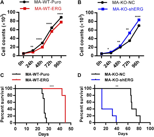
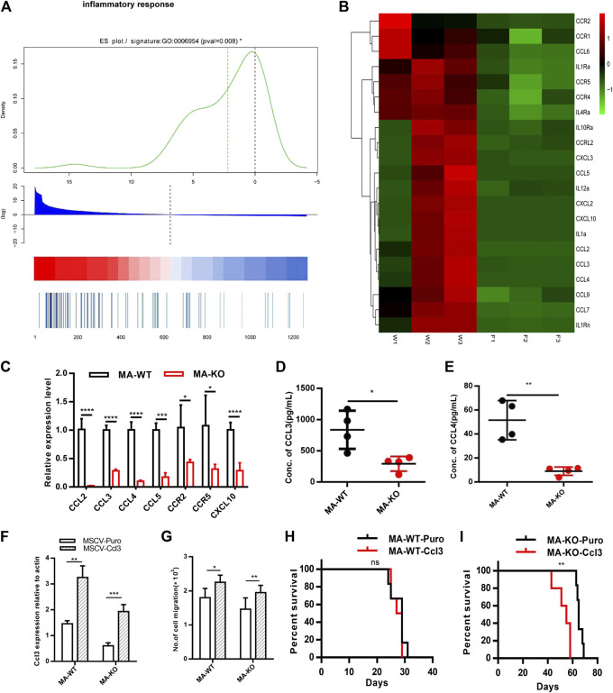
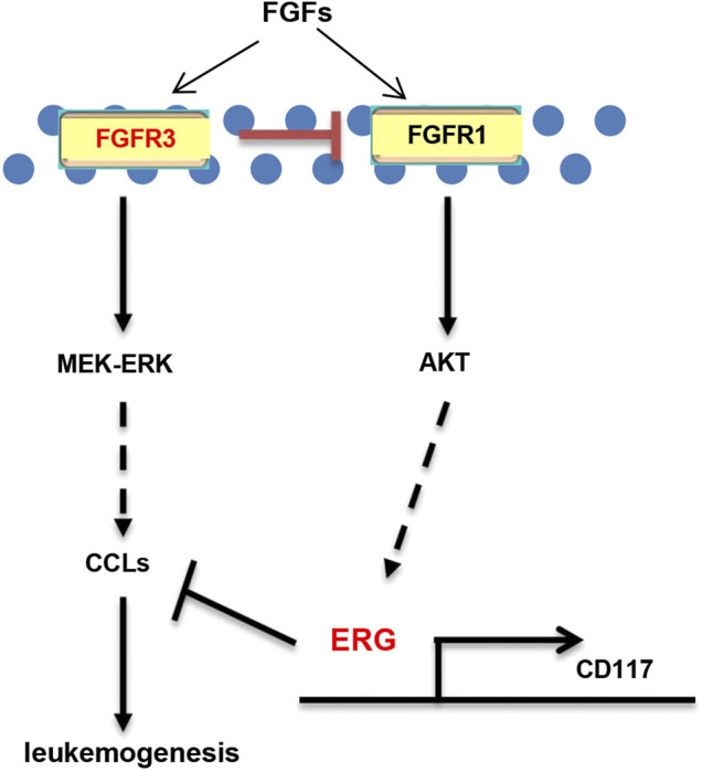
Chemotherapeutic patients with leukemia often relapse and produce drug resistance due to the existence of leukemia stem cells (LSCs). Fibroblast growth factor receptor 3 (FGFR3) signaling mediates the drug resistance of LSCs in chronic myeloid leukemia (CML). However, the function of FGFR3 in acute myeloid leukemia (AML) is less understood. Here, we identified that the loss of FGFR3 reprograms MLL-AF9 (MA)-driven murine AML cells into weakly pathogenic CD117-positive leukemia stem-like cells by activating the FGFR1-ERG signaling pathway. FGFR3 deletion significantly inhibits AML cells engraftment in vivo and extends the survival time of leukemic mice. FGFR3 deletion sharply decreased the expression of chemokines and the prolonged survival time in mice receiving FGFR3-deficient MA cells could be neutralized by overexpression of CCL3. Here we firstly found that FGFR3 had a novel regulatory mechanism for the stemness of LSCs in AML, and provided a promising anti-leukemia approach by interrupting FGFR3.

Keywords: CCL3 chemokine; FGFR1; erg; fibroblast growth factor receptor 3; leukemia (acute myeloid); stem-like cancer cells.


Conflict of interest statement

The authors declare that the research was conducted in the absence of any commercial or financial relationships that could be construed as a potential conflict of interest.

Figures

FIGURE 1
FIGURE 1
Loss of FGFR3 promotes the generation of weakly pathogenic CD117+ MLL-AF9-driven leukemia cells. (A) Representative flow cytometric analysis of MA-WT and MA-KO cells with CD117 and CD11b staining (B) In vitro colony-forming assay of MA-WT and MA-KO cells (n = 3). (C) Growth curve at indicated time from MA-WT and MA-KO cells (n = 5). (D,E) Effects of FGFR3 deletion in MLL-AF9-mediated in vivo leukemogenesis. Kaplan-Meier curves are shown for two groups of transplanted mice including MA-WT (n = 9), and MA-KO (n = 10) in a primary BMT assay (D), and for two groups of transplanted mice including MA-WT (n = 11), and MA-KO (n = 12) in the secondary BMT assay (E).*p < 0.05, **p < 0.01, ***p < 0.001, ****p < 0.0001.
FIGURE 2
FIGURE 2
FGFR3 deletion significantly inhibits in vivo engraftment of leukemic mice. (A–C) The engraftment ability of GFP+ MA-WT and MA-KO cells from BM and SP at day 7 (A), day 14 (B), and day 30 (C) after transplantation. (D) Representative image of hematoxylin and eosin-stained sections at day 30 after transplantation of MA-WT and MA-KO cells. **p < 0.01, ***p < 0.001, ****p < 0.0001.
FIGURE 3
FIGURE 3
RNA-seq and differential gene analysis of MA-WT and MA-KO cells. (A) Heat maps showing gene expression kinetics from RNA-seq results of MA-WT samples (W1, W2, and W3) and MA-KO samples (F1, F2, and F3). (B,C) KEGG enrichment of pathway in MA-KO cells compared to MA-WT cells. (D,E) qRT-PCR assay of differential genes from (A) (n = 4). *p < 0.05, **p < 0.01, ***p < 0.001, ****p < 0.0001.
FIGURE 4
FIGURE 4
FGFR3 deletion programs CD117+ leukemia cells by activating the FGFR1-ERG-CD117 signaling pathway. (A) Representative flow cytometric analysis of CD117+CD11b+/low in MA-WT and MA-KO cells with FGFR inhibitor (PD173074, 4 μM), AKT inhibitor (LY294002, 50 μM), and MEK inhibitor (PD98059, 50 μM). (B) Western blot analysis of ERG expression in 293T cells expressing CA-FGFR1. (C,D) Flow cytometric analysis of CD117+CD11b+/low in WT-ERG cells (C) and KO-shERG cells. (D,E) ChIP-qPCR assay of potential binding of ERG at the promoter region of CD117 (n = 4) (F) luciferase report assay of transcriptional regulation function of ERG (n = 4). (G) Western blot analysis of ERG expression in 293T cells expressing CA-FGFR1 in the presence of signaling inhibitor. (H) The expression of p-FGFR1, p-ERK1/2, p-AKT, and ERG in MA-WT and MA-KO cells treated with PD173074 (5 μM), LY294002 (50 μM), and PD98059 (50 μM) for 16 h **p < 0.01, ****p < 0.0001.
FIGURE 5
FIGURE 5
ERG dosage is essential for the pathogenicity of leukemia cells. (A) The growth curve of MA-WT-ERG cells compared to the corresponding control cells. (B) The growth curve of MA-KO-shERG cells compared to the corresponding control cells. (C) Kaplan-Meier curves are shown for leukemogenesis in two groups of mice transplanted with MA-WT-puro cells (n = 7), and MA-WT-ERG cells (n = 5). (D) Kaplan-Meier curves are shown for leukemogenesis in two groups of mice transplanted with MA-KO-NC cells (n = 5), and MA-KO-shERG cells (n = 5). *p < 0.05, **p < 0.01, ***p < 0.001, ****p < 0.0001.
FIGURE 6
FIGURE 6
FGFR3 deletion decreases the expression of the inflammation factors and the extended survival time of MA-KO cell-transplanted mice could be neutralized by overexpression of CCL3. (A) Gene list rank about inflammatory response genes from RNA-seq data. (B) Heat map on the expression level of inflammatory response genes in MA-WT samples (W1, W2, and W3) and MA-KO samples (F1, F2, and F3). (C) Differential expression genes from (B) by qRT-PCR analysis (n = 4). (D,E) The concentration of CCL3 (D) and CCL4 (E) in BM supernatant of leukemic mice at day 30 after transplantation by ELISA (n = 4). (F) Overexpression of CCL3 in MA-WT and MA-KO cells (n = 4). (G) Number of cell migration from 100,000MA-WT-CCL3 or MA-KO-CCL3 cells compared with respective control cells (n = 3). (H,I) Kaplan-Meier curves are shown for in vivo leukemogenesis in two groups of mice transplanted with leukemia cells, including MA-WT-puro cells (n = 6), and MA-WT-CCL3 cells (n = 5) (H) as well as MA-KO-puro cells (n = 6), and MA-KO-CCL3 cells (n = 5) (I). *p < 0.05, **p < 0.01, ***p < 0.001, ****p < 0.0001.
FIGURE 7
FIGURE 7
Schematic model of FGFR3 signaling in AML. Schematic model showed that FGFR3 might upregulate CCL expression by activating MEK-ERK1/2 to maintain leukemogenesis. In addition, FGFR3 deletion programs weakly pathogenic CD117+ leukemia stem-like cells by activating FGFR1-AKT-ERG-CD117 signaling. Furthermore, ERG does not only promote the stemness of LSCs by upregulating the expression of CD117, but also suppresses in vivo leukemogenesis by inhibiting the expression of chemokines genes.

Similar articles

Cited by

References

    1. Baba T., Naka K., Morishita S., Komatsu N., Hirao A., Mukaida N. (2013). MIP-1α/CCL3-mediated maintenance of leukemia-initiating cells in the initiation process of chronic myeloid leukemia. J. Exp. Med. 210 (12), 2661–2673. 10.1084/jem.20130112 - DOI - PMC - PubMed
    1. Baba T., Tanabe Y., Yoshikawa S., Yamanishi Y., Morishita S., Komatsu N., et al. (2016). MIP-1α/CCL3-expressing basophil-lineage cells drive the leukemic hematopoiesis of chronic myeloid leukemia in mice. Blood 127 (21), 2607–2617. 10.1182/blood-2015-10-673087 - DOI - PubMed
    1. Batta K., Florkowska M., Kouskoff V., Lacaud G. (2014). Direct reprogramming of murine fibroblasts to hematopoietic progenitor cells. Cell Rep. 9 (5), 1871–1884. 10.1016/j.celrep.2014.11.002 - DOI - PMC - PubMed
    1. de Haan G., Weersing E., Dontje B., van Os R., Bystrykh L. V., Vellenga E., et al. (2003). In vitro generation of long-term repopulating hematopoietic stem cells by fibroblast growth factor-1. Dev. Cell 4 (2), 241–251. 10.1016/s1534-5807(03)00018-2 - DOI - PubMed
    1. Deng C., Wynshaw-Boris A., Zhou F., Kuo A., Leder P. (1996). Fibroblast growth factor receptor 3 is a negative regulator of bone growth. Cell 84 (6), 911–921. 10.1016/s0092-8674(00)81069-7 - DOI - PubMed

LinkOut - more resources