. 2021 Feb 1:12:611984.
doi: 10.3389/fphys.2021.611984. eCollection 2021.

A Longitudinal Pilot Study on Cognition and Cerebral Hemodynamics in a Mouse Model of Preeclampsia Superimposed on Hypertension: Looking at Mothers and Their Offspring

Affiliations

A Longitudinal Pilot Study on Cognition and Cerebral Hemodynamics in a Mouse Model of Preeclampsia Superimposed on Hypertension: Looking at Mothers and Their Offspring

Lianne J Trigiani et al. Front Physiol. .

Abstract

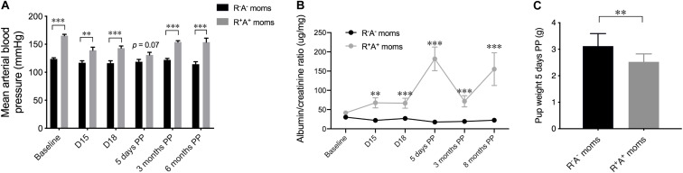
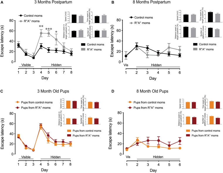
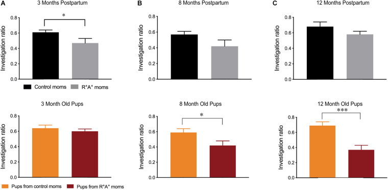
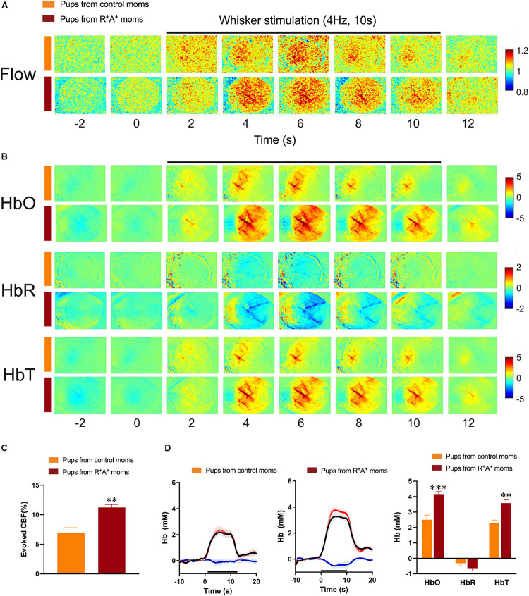
Preeclampsia is a common hypertensive disorder in pregnant women and whose causes and consequences have focused primarily on cardiovascular outcomes on the mother and offspring, often without taking into consideration the possible effects on the brain. One possible cause of preeclampsia has been attributed to alterations in the renin-angiotensin system, which has also been linked to cognitive decline. In this pilot study, we use a transgenic mouse model that chronically overexpresses human angiotensinogen and renin (R+A+ mice) that displayed characteristics of preeclampsia such as proteinuria during gestation. Offspring of these mothers as well as from control mothers were also examined. We were primarily interested in detecting whether cognitive deficits were present in the mothers and offspring in the long term and used a spatial learning and memory task as well as an object recognition task at three timepoints: 3, 8, and 12 months post-partum or post-natal, while measuring blood pressure and performing urine analysis after each timepoint. While we did not find significant deficits in preeclamptic mothers at the later timepoints, we did observe negative consequences in the pups of R+A+ mice that coincided with hemodynamic alterations whereby pups had higher whisker-evoked oxygenated hemoglobin levels and increased cerebral blood flow responses compared to control pups. Our study provides validation of this preeclampsia mouse model for future studies to decipher the underlying mechanisms of long-term cognitive deficits found in offspring.

Keywords: cerebral hemodynamics; cerebrovascular function; cognition; preeclampsia; renin—angiotensin—aldosterone system.


Conflict of interest statement

The authors declare that the research was conducted in the absence of any commercial or financial relationships that could be construed as a potential conflict of interest.

Figures

FIGURE 1
FIGURE 1
Preeclampsia model characteristics. (A) Mean arterial blood pressure shown at different timepoints for the mothers throughout the study beginning at baseline prior to mating, two times during pregnancy and three postpartum (PP) timepoints, revealing that this model is chronically hypertensive. (B) Urinalysis quantification over the same timepoints in mothers revealing an increased albumin:creatine ratio in R+A+ mothers during pregnancy that persisted up to 8 months PP. (C) The average pup weight 5 days PP from individual mothers was averaged and shown here indicating R+A+ birthed smaller pups than control mothers. N = 10–13 for control mothers and n = 5–8 for preeclamptic (R+A+) mothers, **p < 0.01 and ***p < 0.001.
FIGURE 2
FIGURE 2
Spatial learning and memory testing in mothers and offspring. (A) Spatial learning portion of MWM in mothers 3 months PP showing deficits in preeclamptic mothers (R+A+, n = 7) early during the learning phase but without any spatial memory deficits measured during the probe trial compared to control mothers (n = 12). (C) Regardless of the genotype of their birth mother, 3-months old pups (n = 18–26/group) showed no differences in spatial learning nor memory. (B) At 8 months PP another MWM was conducted, revealing no spatial learning and memory deficits in the mothers nor in the pups (D). Sample size at 8 months PP, control mothers n = 10, R+A+ mothers n = 6. Due to no observed sex differences in pups at 3 months of age, group sizes were reduced at random when tested at 8 months to n = 11/group. *p < 0.05, **p < 0.01, and ***p < 0.001.
FIGURE 3
FIGURE 3
Novel object recognition memory testing in mothers and offspring. (A) NOR investigation ratios for mothers 3 months PP (R+A+ n = 7, controls n = 12) and pups at 3 months of age (n = 18–26/group) revealing a deficit in preeclamptic mothers. While no deficits were observed in mothers at 8 (B) nor 12 months PP (C), pups born from preeclamptic mothers had impaired performance at these two age points (B,C). Samples sizes at 8 and 12 months: control mothers n = 10, R+A+ mothers n = 5–6, pups n = 11/group. *p < 0.05 ***p < 0.001.
FIGURE 4
FIGURE 4
Evaluation of hemodynamic alterations in offspring at 12 months of age. (A) Functional laser speckle contrast imaging maps representative of cerebral blood flow (CBF) changes in the barrel cortex in response to whisker stimulation. (B) Functional imaging maps of oxygenated (HbO), deoxygenated (HbR) and total hemoglobin (HbT) levels from 4 s prior to stimulus to 2 s after a 10 s whisker stimulation (4 Hz) in offspring of preeclamptic (red bars) and control mothers (orange bars). (C) Quantification of laser Speckle contrast imaging showing an increased evoked CBF response in pups from preeclamptic mothers. (D) Average traces and quantification of peak concentrations (mM) of HbO (red line), HbR (blue line), and HbT (black line) from offspring revealing a significant increase in HbO and HbT levels in pups from preeclamptic mothers. N = 6/group **p < 0.01 and ***p < 0.001.

Similar articles

Cited by

References

    1. Apicella C., Ruano C. S. M., Méhats C., Miralles F., Vaiman D. (2019). The role of epigenetics in placental development and the etiology of preeclampsia. Int. J. Mol. Sci. 20:2837. 10.3390/ijms20112837 - DOI - PMC - PubMed
    1. Belfort M. A., Grunewald C., Saade G. R., Varner M., Nisell H. (1999). Preeclampsia may cause both overperfusion and underperfusion of the brain. Acta Obstet. Gynecol. Scand. 78 586–591. 10.1034/j.1600-0412.1999.780705.x - DOI - PubMed
    1. Berks D., Steegers E. A. P., Molas M., Visser W. (2009). Resolution of hypertension and proteinuria after preeclampsia. Obstet. Gynecol. 114 1307–1311. 10.1097/aog.0b013e3181c14e3e - DOI - PubMed
    1. Bokslag A., van Weissenbruch M., Mol B. W., de Groot C. J. (2016). Preeclampsia; short and long-term consequences for mother and neonate. Early Hum. Dev. 102 47–50. - PubMed
    1. Chen B. R., Kozberg M. G., Bouchard M. B., Shaik M. A., Hillman E. M. (2014). A critical role for the vascular endothelium in functional neurovascular coupling in the brain. J. Am. Heart Assoc. 3:e000787. - PMC - PubMed

LinkOut - more resources