

Impaired Ganglion Cell Function Objectively Assessed by the Photopic Negative Response in Affected and Asymptomatic Members From Brazilian Families With Leber's Hereditary Optic Neuropathy
- PMID: 33584522
- PMCID: PMC7874135
- DOI: 10.3389/fneur.2020.628014
Impaired Ganglion Cell Function Objectively Assessed by the Photopic Negative Response in Affected and Asymptomatic Members From Brazilian Families With Leber's Hereditary Optic Neuropathy
Abstract
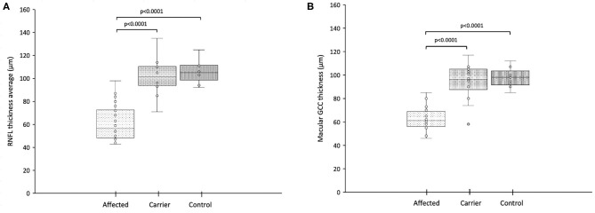
Purpose: The photopic negative response (PhNR) is an electrophysiological method that provides retinal ganglion cell function assessment using full-field stimulation that does not require clear optics or refractive correction. The purpose of this study was to assess ganglion cell function by PhNR in affected and asymptomatic carriers from Brazilian families with LHON. Methods: Individuals either under suspicion or previously diagnosed with LHON and their family members were invited to participate in this cross-sectional study. Screening for the most frequent LHON mtDNA mutations was performed. Visual acuity, color discrimination, visual fields, pattern-reversal visual evoked potentials (PRVEP), full-field electroretinography and PhNR were tested. A control group of healthy subjects was included. Full-field ERG PhNR were recorded using red (640 nm) flashes at 1 cd.s/m2, on blue (470 nm) rod saturating background. PhNR amplitude (μV) was measured using baseline-to-trough (BT). Optical coherence tomography scans of both the retinal nerve fiber layer (RNFL) and ganglion cell complex (GCC) were measured. PhNR amplitudes among affected, carriers and controls were compared by Kruskal-Wallis test followed by post-hoc Dunn test. The associations between PhNR amplitude and OCT parameters were analyzed by Spearman rank correlation. Results: Participants were 24 LHON affected patients (23 males, mean age=30.5 ± 11.4 yrs) from 19 families with the following genotype: m.11778G>A [N = 15 (62%), 14 males]; m.14484T>C [N = 5 (21%), all males] and m.3460G>A [N = 4 (17%), all males] and 14 carriers [13 females, mean age: 43.2 ± 13.3 yrs; m.11778G>A (N = 11); m.3460G>A (N = 2) and m.14484T>C (N = 1)]. Controls were eight females and seven males (mean age: 32.6 ± 11.5 yrs). PhNR amplitudes were significantly reduced (p = 0.0001) in LHON affected (-5.96 ± 3.37 μV) compared to carriers (-16.53 ± 3.40 μV) and controls (-23.91 ± 4.83; p < 0.0001) and in carriers compared to controls (p = 0.01). A significant negative correlation was found between PhNR amplitude and total macular ganglion cell thickness (r = -0.62, p < 0.05). Severe abnormalities in color discrimination, visual fields and PRVEPs were found in affected and subclinical abnormalities in carriers. Conclusions: In this cohort of Brazilian families with LHON the photopic negative response was severely reduced in affected patients and mildly reduced in asymptomatic carriers suggesting possible subclinical abnormalities in the latter. These findings were similar among pathogenic mutations.
Keywords: electroretinography; leber's hereditary optic neuropathy; photopic negative response; retinal ganglion cell; visual evoked cortical potentials.
Copyright © 2021 Botelho, Salomão, Tengan, Karanjia, Moura, Rocha, Silva, Fernandes, Watanabe, Sacai, Belfort, Carelli, Sadun and Berezovsky.
Conflict of interest statement
The authors declare that the research was conducted in the absence of any commercial or financial relationships that could be construed as a potential conflict of interest.
Figures


References
-
- Barboni P, Sadun AA, Balducci N, Carelli V. Natural history. Atlas of LHON. In: Barboni P, Sadun AA, editors. Chapter I. Netherlands: MEDonline International; (2019). p. 6–29.
LinkOut - more resources
Full Text Sources
Other Literature Sources