. 2021 Jan 28:11:583229.
doi: 10.3389/fendo.2020.583229. eCollection 2020.

Additive Effects of VDBP and 1,25(OH)2D3 on the Viability and Apoptosis of Rheumatoid Arthritis Synovial Fibroblasts

Affiliations

Additive Effects of VDBP and 1,25(OH)2D3 on the Viability and Apoptosis of Rheumatoid Arthritis Synovial Fibroblasts

Yeyong Zhang et al. Front Endocrinol (Lausanne). .

Abstract

Aim: This study is to investigate the additive effect of Vitamin D-binding protein (VDBP) and 1,25(OH)2D3 on the viability and apoptosis of synovial cells from patients with rheumatoid arthritis (RA).

Methods: Synovial tissues and synovial fluid of patients with RA and osteoarthritis (OA) were collected. The expression of VDBP was analyzed with immunohistochemistry and ELISA. CCK-8 assay was applied to detect cell viability. Flow cytometry was used to analyze cell cycle and apoptosis.

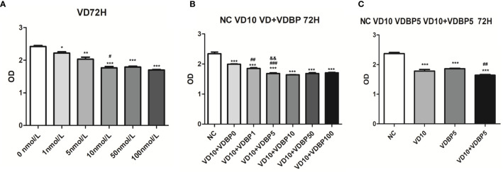
Results: Immunohistochemical results showed that the expression of VDBP in the synovium of RA patients was significantly lower than that of OA (P<0.05). Similarly, ELISA results presented a lower expression of VDBP in the synovial fluid of RA patients. The results of CCK-8 assay showed that both 1,25(OH)2D3 and VDBP significantly inhibited the viability of rheumatoid arthritis synovial fibroblasts (RASF) (P<0.05). The treatment with 1,25(OH)2D3+VDBP led to more significantly inhibited viability of RASF, compared with 1,25(OH)2D3 alone (P<0.05). The results of flow cytometry showed that 1,25(OH)2D3 and VDBP both promoted the apoptosis of RASF (P<0.05) and 1,25(OH)2D3+VDBP led to a higher proportion of RASF apoptosis, compared with 1,25(OH)2D3 alone (P<0.05). However, 1,25(OH)2D3 and VDBP had no significant effect on the cell cycle of RASF. Additionally, 1,25(OH)2D3 promoted the expression of VDBP in RASF, but not concentration-dependently.

Conclusion: VDBP is reduced in the synovial tissue and synovial fluid of RA patients and can inhibit viability of RASF and promote the apoptosis of RASF. The 1,25(OH)2D3 can upregulate the expression of VDBP in RASF. Additionally, VDBP can enhance the effects of 1,25(OH)2D3 on viability and apoptosis of RASF.

Keywords: 1,25(OH)2D3; apoptosis; proliferation; rheumatoid arthritis synovial fibroblast; vitamin D-binding protein.


Conflict of interest statement

The authors declare that the research was conducted in the absence of any commercial or financial relationships that could be construed as a potential conflict of interest.

Figures

Figure 1
Figure 1
Analysis of Vitamin D-binding protein (VDBP) expression. (A) Expression of VDBP in synovium of osteoarthritis (OA) and rheumatoid arthritis (RA) patients. Immunohistochemistry was performed to detect VDBP expression. Representative immunohistochemistry results (at 200 x and 400 x magnification) and relative level of VDBP were shown. (B) The level of VDBP in synovial fluid of RA and OA patients. Enzyme-linked immunosorbent assay (ELISA) was conducted to detect VDBP in synovial fluid. (C) VDBP protein level in OASF and rheumatoid arthritis synovial fibroblasts (RASF). Western blot was carried out to detect protein expression in three RA patients and three OA patients. The representative Western blot results and the relative expression of VDBP from western blot were shown. Each lane represents the sample from an individual patient. Several western blots were used for quantification. *P < 0.05, **P < 0.01, ***P < 0.001.
Figure 2
Figure 2
Effect of Vitamin D-binding protein (VDBP) on viability, apoptosis and cell cycle of RASF. (A) Effect of VDBP on viability of rheumatoid arthritis synovial fibroblasts (RASF). Human recombinant VDBP (0, 1, 5, 10, 50, 100 ng/ml) was added to RASF for 72 h. Cell viability was assessed with CCK-8 assay. Compared with 0 ng/ml, **P < 0.01, ***P < 0.001. Compared with 1 ng/ml, #P < 0.05. (B) Apoptosis analysis of RASF cells with or without 5 ng/ml VDBP treatment. RASF cells were treated without (NC) or with 5 ng/ml VDBP for 72 h. Flow cytometry was used to detect apoptosis. Representative and quantitative flow cytometry results were shown. ***P < 0.001. (C) Cell cycle analysis of RASF cells treated with or without 5 ng/ml VDBP. RASF cells were treated without (NC) or with 5 ng/ml VDBP for 72 h. Flow cytometry was used to detect cell cycle. Representative and quantitative flow cytometry results were shown.
Figure 3
Figure 3
Effect of 1,25(OH)2D3, Vitamin D-binding protein (VDBP), and 1,25(OH)2D3+VDBP on RASF. (A) The effect of 1,25(OH)2D3 on the viability of rheumatoid arthritis synovial fibroblasts (RASF) cells. RASF cells were treated with different concentrations of 1,25(OH)2D3 (0,1,5,10,50,100 nmol/L) for 72 h. Cell viability was assessed with CCK-8 assay. Compared with 0 nmol/L, *P < 0.05, **P < 0.01, ***P < 0.001. Compared with 5 nmol/L, #P < 0.05. (B) The enhanced inhibition of 1,25(OH)2D3 with VDBP on the viability of RASF. RASF cells were treated with 1,25(OH)2D3+VDBP as indicated in the figure. Compared with NC, ***P<0.001. Compared with VD10+VDBP0, ##P < 0.01, ###P < 0.001. Compared with VD10+VDBP1, &&P<0.01. (C) The enhanced inhibition of 1,25(OH)2D3 with VDBP on the viability of RASF. RASF cells were treated with 1,25(OH)2D3, VDBP, and 1,25(OH)2D3+VDBP as indicated in the figure. Compared with NC, ***P < 0.001. Compared with VD10, ##P < 0.01.
Figure 4
Figure 4
Flow cytometry analysis of apoptosis and cell cycle. (A) Apoptosis analysis of rheumatoid arthritis synovial fibroblasts (RASF) after treatment with 1,25(OH)2D3, VDBP and 1,25(OH)2D3+VDBP. RASF cell treatment was indicated in the figure. Flow cytometry was used to detect apoptosis. Representative and quantitative flow cytometry results were shown. *P<0.05, **P<0.01, ***P<0.001. (B) Cell cycle analysis of RASF after treatment with 1,25(OH)2D3, VDBP and 1,25(OH)2D3+VDBP. RASF cell treatment was indicated in the figure. Flow cytometry was used to detect cell cycle. Representative and quantitative flow cytometry results were shown. ns, not significant.
Figure 5
Figure 5
Effect of 1,25(OH)2D3 on the expression of Vitamin D-binding protein (VDBP). Rheumatoid arthritis synovial fibroblasts (RASF) was treated with different concentrations of 1,25(OH)2D3. Western blot was used to detect VDBP protein expression. The representative Western blot results and the relative expression of VDBP from western blot were shown. **P < 0.01. ns, not significant.

Similar articles

Cited by

References

    1. Svensson B, Andersson M, Forslind K, Ajeganova S, Hafström I. Persistently active disease is common in patients with rheumatoid arthritis, particularly in women: a long-term inception cohort study. Scand J Rheumatol (2016) 45:448–55. 10.3109/03009742.2016.1147595 - DOI - PubMed
    1. Tsuprykov O, Chen X, Hocher CF, Skoblo R, Lianghong Y, Hocher B. Why should we measure free 25(OH) vitamin D? J Steroid Biochem Mol Biol (2018) 180:87–104. 10.1016/j.jsbmb.2017.11.014 - DOI - PubMed
    1. Mukherjee D, Lahiry S, Thakur S, Chakraborty DS. Effect of 1,25 dihydroxy vitamin D3 supplementation on pain relief in early rheumatoid arthritis. J Family Med Prim Care (2019) 8:517–22. 10.4103/jfmpc.jfmpc_446_18 - DOI - PMC - PubMed
    1. Tanaka S, Suto A, Iwamoto T, Kashiwakuma D, Kagami S, Suzuki K, et al. Sox5 and c-Maf cooperatively induce Th17 cell differentiation via RORγt induction as downstream targets of Stat3. J Exp Med (2014) 211:1857–74. 10.1084/jem.20130791 - DOI - PMC - PubMed
    1. Zakeri Z, Sandoughi M, Mashhadi MA, Raeesi V, Shahbakhsh S. Serum vitamin D level and disease activity in patients with recent onset rheumatoid arthritis. Int J Rheum Dis (2016) 19:343–7. 10.1111/1756-185X.12181 - DOI - PubMed

Publication types

MeSH terms