Review
. 2021 Jan 27:11:619352.
doi: 10.3389/fendo.2020.619352. eCollection 2020.

Functional Pituitary Networks in Vertebrates

Affiliations
Review

Functional Pituitary Networks in Vertebrates

Yorgui Santiago-Andres et al. Front Endocrinol (Lausanne). .

Abstract

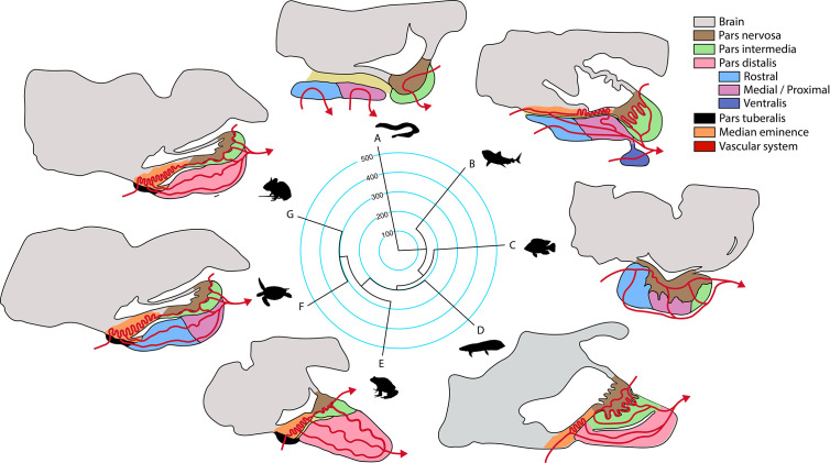
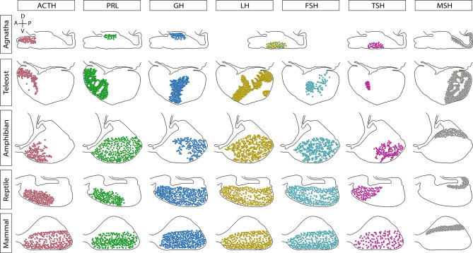
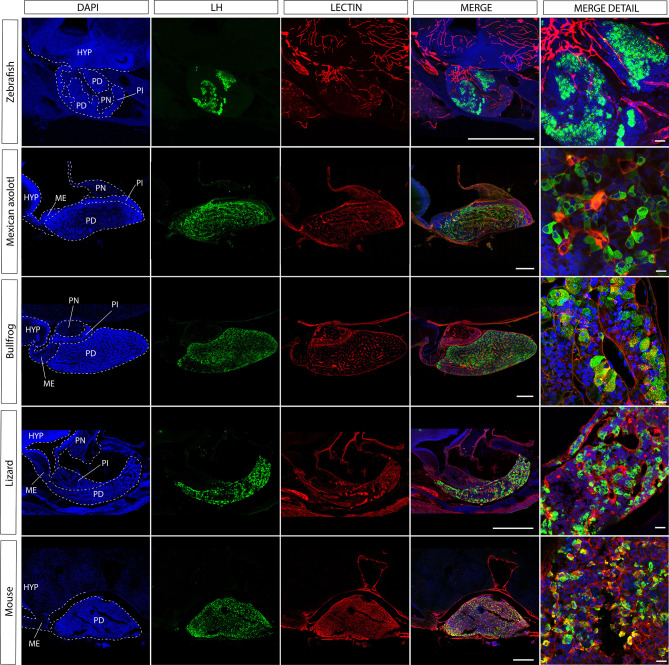
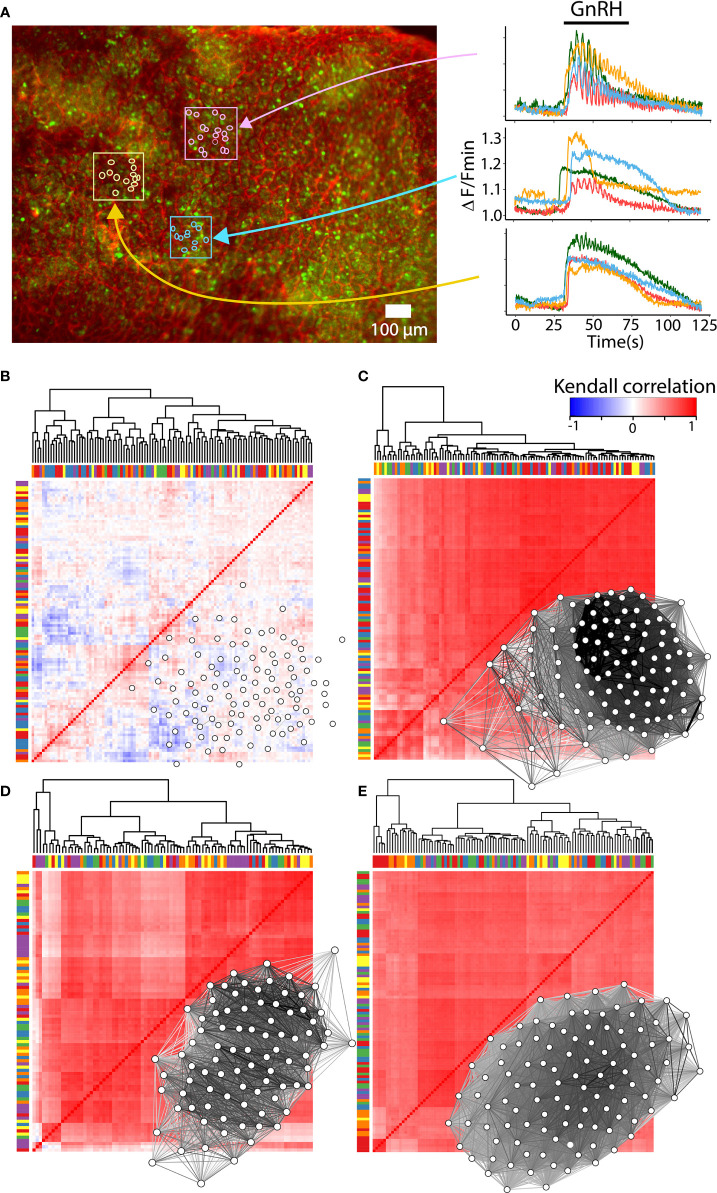
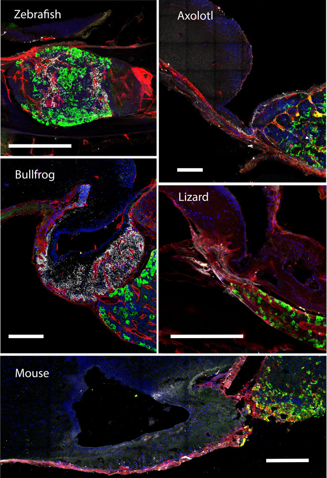
The pituitary is a master endocrine gland that developed early in vertebrate evolution and therefore exists in all modern vertebrate classes. The last decade has transformed our view of this key organ. Traditionally, the pituitary has been viewed as a randomly organized collection of cells that respond to hypothalamic stimuli by secreting their content. However, recent studies have established that pituitary cells are organized in tightly wired large-scale networks that communicate with each other in both homo and heterotypic manners, allowing the gland to quickly adapt to changing physiological demands. These networks functionally decode and integrate the hypothalamic and systemic stimuli and serve to optimize the pituitary output into the generation of physiologically meaningful hormone pulses. The development of 3D imaging methods and transgenic models have allowed us to expand the research of functional pituitary networks into several vertebrate classes. Here we review the establishment of pituitary cell networks throughout vertebrate evolution and highlight the main perspectives and future directions needed to decipher the way by which pituitary networks serve to generate hormone pulses in vertebrates.

Keywords: evolution; networks; pituitary; plasticity; vertebrates.


Conflict of interest statement

The authors declare that the research was conducted in the absence of any commercial or financial relationships that could be construed as a potential conflict of interest.

Figures

Figure 1
Figure 1
Phylogeny of the principal groups of vertebrates and anatomical pituitary organization (anterior-caudal-dorso-ventral orientation). Terminal branches of the phylogenetic tree show a representative organism of each clade (A, jawless fish; B, cartilaginous fish; C, ray-finned fish; D, lungfish; E, amphibian; F, reptile that includes birds; G, mammal) and their sagittal representation of the main structures in the pituitary gland according to (23). Median divergence times from the TimeTree database were used to construct the tree, numbers in the phylogeny are million years.
Figure 2
Figure 2
Sagittal representation of the endocrine cells distribution in the pituitary gland of vertebrates. The distribution of cell types has been modified over the evolution of vertebrates. In agnathans and teleosts, here represented by the sea lamprey and Nile tilapia, respectively, cells are markedly regionalized. Amphibians and reptiles present a regionalized distribution of some cell types, but others are scattered through the pars distalis. In contrast, the mammalian secreting cells are widely scattered throughout the lobe. In lampreys there is only one cell type producing glycoprotein hormone (GPH, a gonadotropin homologue) and thyrostimulin (TSN, a TSH homologue). Additionally, teleost fish possess a unique cell type that produces somatolactin, the somato-lactotroph, not shown. A, anterior or rostral; P, posterior or caudal; D, dorsal; V, ventral.
Figure 3
Figure 3
The gonadotroph network and the relation with the hypophyseal portal system in vertebrates. Midsagittal view of the pituitary gland in relation to the hypothalamus (anterior at the left, dorsal to the top) using confocal microscopy. Shown are immunostainings for LHβ (green) in five different species of vertebrates (zebra fish, Danio rerio; axolotl, Ambystoma mexicanum; bullfrog, Lithobates catesbeianus; lizard, Sceloporus aeneus; mouse, Mus musculus). In contrast to tetrapods, where LH-containing gonadotrophs are well distributed in the pars distalis, in teleost these cells are confined to the rostral pars distalis. The merge detail column is a power magnification of the precedent, showing that gonadotrophs establish cell-cell contact to form an interdigitated network and reveal that this pattern of cell organization is extended to all vertebrates. Note that the vascular network pervades the pars distalis and gonadotrophs form process to the capillaries (red, lectin-rhodamine), with the exception of zebrafish. DAPI counterstain is shown in blue. Dashed lines mark the three main regions of the pituitary. HYP, hypothalamus; PD, pars distalis; PI, pars intermedia; PN, pars nervosa; ME, median eminence. Scale bars represent 400 μm in whole-gland views and 20 μm for merge detail.
Figure 4
Figure 4
Reconfiguration of the gonadotroph network throughout the reproductive cycle. (A) Intracellular calcium recordings in ex vivo preparations of mouse pituitary loaded with the calcium sensor Fluo 4-AM (green) and rhodamine for vasculature (red). Monitoring of calcium activity was performed on the ventral side of the gland under an epifluorescence stereomicroscope and continuously perfused with Ringer’s solution [methods as in (146)]. Color traces show the intracellular calcium activity of individual gonadotrophs in response to 10mM GnRH, indicating clusters of gonadotrophs surrounded by vasculature with a similar (pink and yellow boxes), or heterogenous (blue box) calcium response. (B) Heatmap and network organization of basal calcium activity of correlated pairs of gonadotropes from a male mouse. Gonadotrophs stimulated with GnRH in male mouse (C), female mouse in diestrus (D), and proestrus (E) showed an increase in synchronized calcium activity compared to basal activity. Notice that the overall correlation values are similar between diestrus and proestrus, as well as their connectivity. Bars in the heatmaps indicate sorting of gonadotropes by correlation but not by spatial proximity, while groups of gonadotrophs surrounded by vasculature are represented with the same color in bars. Network maps were plotted using qgraph and significant pairs (edges) of correlated cells (nodes) are shown (P < 0.05).
Figure 5
Figure 5
A multimodal communication between the hypothalamus and pituitary cell networks. Shown are sagittal sections (20 μm) of 4% paraformaldehyde fixed tissue, anterior at the left, dorsal to the top. Immunocytochemistry for gonadotrophs containing the LHβ subunit (green) and GnRH neurons (gray), species as in Figure 3 . The distribution of gonadotrophs in the pars distalis, proximal to the hypothalamus, have remarkable cell-cell contacts in the tetrapod species, in teleost these cells are exclusively found in the rostral pars distalis forming a clear-cut cluster. The gonadotroph network arrangement is highly influenced by the vascular system (red; lectin-rhodamine), since these cells have cytoplasmic extensions to the capillaries. In tetrapods, the GnRH-producing neurons extend their projections to the median eminence where GnRH is secreted to the capillaries. In contrast, the GnRH neurons in teleost unfold throughout the pituitary and establish at the boundaries of the gonadotroph patch, following the vascular system. Note the close proximity between gonadotrophs and GnRH at the median eminence of tetrapods. DAPI is shown in blue for counterstain. Scale bars represent 200 μm.

Similar articles

Cited by

References

    1. West-Eberhard MJ. Developmental plasticity and the origin of species differences. Proc Natl Acad Sci USA (2005) 102 Suppl 1:6543–9. 10.1073/pnas.0501844102 - DOI - PMC - PubMed
    1. Le Tissier P, Campos P, Lafont C, Romanò N, Hodson DJ, Mollard P. An updated view of hypothalamic-vascular-pituitary unit function and plasticity. Nat Rev Endocrinol (2017) 13:257–67. 10.1038/nrendo.2016.193 - DOI - PubMed
    1. Schlosser G. From so simple a beginning – what amphioxus can teach us about placode evolution. Int J Dev Biol (2017) 61:633–48. 10.1387/ijdb.170127gs - DOI - PubMed
    1. Rathke H. Ueber die Entstehung der Glandula pituitaria. Arch Anat Physiol Sci Med (1838) 40:482–5.
    1. Fabian P, Tseng K-C, Smeeton J, Lancman JJ, Duc Si Dong P, Cerny R, et al. Lineage analysis reveals an endodermal contribution to the vertebrate pituitary. Science (2020) 370:463–7. 10.1126/science.aba4767 - DOI - PMC - PubMed

Publication types

LinkOut - more resources