. 2021 Jan 15:11:611122.
doi: 10.3389/fmicb.2020.611122. eCollection 2020.

Rv1717 Is a Cell Wall - Associated β-Galactosidase of Mycobacterium tuberculosis That Is Involved in Biofilm Dispersion

Affiliations

Rv1717 Is a Cell Wall - Associated β-Galactosidase of Mycobacterium tuberculosis That Is Involved in Biofilm Dispersion

Suman Bharti et al. Front Microbiol. .

Abstract

Understanding the function of conserved hypothetical protein (CHP)s expressed by a pathogen in the infected host can lead to better understanding of its pathogenesis. The present work describes the functional characterization of a CHP, Rv1717 of Mycobacterium tuberculosis (Mtb). Rv1717 has been previously reported to be upregulated in TB patient lungs. Rv1717 belongs to the cupin superfamily of functionally diverse proteins, several of them being carbohydrate handling proteins. Bioinformatic analysis of the amino acid sequence revealed similarity to glycosyl hydrolases. Enzymatic studies with recombinant Rv1717 purified from Escherichia coli showed that the protein is a β-D-galactosidase specific for pyranose form rather than the furanose form. We expressed the protein in Mycobacterium smegmatis (Msm), which lacks its ortholog. In Msm Rv1717 , the protein was found to localize to the cell wall (CW) with a preference to the poles. Msm Rv1717 showed significant changes in colony morphology and cell surface properties. Most striking observation was its unusual Congo red colony morphotype, reduced ability to form biofilms, pellicles and autoagglutinate. Exogenous Rv1717 not only prevented biofilm formation in Msm, but also degraded preformed biofilms, suggesting that its substrate likely exists in the exopolysaccharides of the biofilm matrix. Presence of galactose in the extracellular polymeric substance (EPS) has not been reported before and hence we used the galactose-specific Wisteria floribunda lectin (WFL) to test the same. The lectin extensively bound to Msm and Mtb EPS, but not the bacterium per se. Purified Rv1717 also hydrolyzed exopolysaccharides extracted from Msm biofilm. Eventually, to decipher its role in Mtb, we downregulated its expression and demonstrate that the strain is unable to disperse from in vitro biofilms, unlike the wild type. Biofilms exposed to carbon starvation showed a sudden upregulation of Rv1717 transcripts supporting the potential role of Rv1717 in Mtb dispersing from a deteriorating biofilm.

Keywords: biofilm; biofilm dispersion; extracellular polymeric substance; galactosidase; mycobacteria.


Conflict of interest statement

The authors declare that the research was conducted in the absence of any commercial or financial relationships that could be construed as a potential conflict of interest.

Figures

Figure 1
Figure 1
Recombinant Rv1717 purified from Escherichia coli shows β-galactosidase activity. (A) SDS-PAGE of His-tagged Rv1717 protein (13.7 kDa) purified by Ni-nitrilotriacetic acid (Ni-NTA) affinity chromatography. (B) Glycosyl hydrolase activity of Rv1717 was tested using a set of p-nitrophenyl (pNP)-glycosides, pNP-β-D-glucopyranoside (pNP-β-D-Glu), pNP-β-D-galactopyranoside (pNP-β-D-Gal), pNP-β-D-mannopyranoside (pNP-β-D-Man), pNP-α-L-arabinopyranoside (pNP-α-L-Ara), pNP-α-L-rhamnopyranoside (pNP-α-L-Rha), pNP-β-D-glucopyranosiduronic acid (pNP-β-D-GlcA), pNP-α-D-galactopyranoside (pNP-α-D-Gal), pNP-β-D-galactofuranoside (pNP-β-D-Galf), and pNP-2-acetamido-2-deoxy-β-D-galactopyranoside (pNP-β-D-GalNAc) as substrates. One unit (U) of enzyme activity was defined as the amount of enzyme that released 1 μmol of p-nitrophenol (pNP) from pNP-glycosides per minute under the standard assay condition. Graph shows the enzymatic activity of Rv1717 obtained for various substrates relative to the buffer without the protein. Inset shows the standard curve of pNP used to calculate the enzymatic release of pNP. (C,D) The effect of pH and temperature respectively, on the enzymatic activity with pNP-β-D-Gal substrate. (E) Effect of adding metal ions or EDTA (final concentration of 0.5 mM) to the enzymatic reaction with pNP-β-D-Gal substrate. Control reaction contains none of these. Data plotted are mean ± SD of three independent experiments.
Figure 2
Figure 2
Mycobacterium smegmatis expressing Rv1717 shows altered colony morphology and cell wall (CW) associated properties. (A) On MB 7H10 agar, M. smegmatis harboring empty vector (MsmpMV261) formed rough colonies with irregular margins, while M. smegmatis expressing Rv1717 (MsmRv1717) formed smoother colonies with more regular margins. (B) Accumulation of EtBr and (C) Nile red by MsmpMV261 and MsmRv1717 (D) MsmRv1717 and MsmpMV261 were left untreated or treated with 0.05 or 0.1% (w/v) Sodium dodecyl sulfate (SDS) for 3 and 6 h. Viable cells in all samples were estimated by CFU assay. Statistical significance of data wherever applicable is indicated by ns: p > 0.05; ***p < 0.001. Data plotted are mean ± SD of three independent experiments.
Figure 3
Figure 3
Rv1717 expressed in M. smegmatis localizes to the CW and prefers the poles. (A) Western blot analysis of equal amounts (30 μg protein) of whole cell lysate (WCL), CW, cell membrane (CM), and cytosolic (CY) fractions of MsmRv1717. Rv1717 which was expressed with a C-terminal 6 × His tag was detected by anti-6 × His tag antibody. Lipoarabinomannan (LAM) and Hsp65 were detected using anti-M. smegmatis LAM monoclonal antibody NR-13798 (BEI Resources, Manassas, VA) and anti-Hsp65 monoclonal antibody, respectively. LAM was used as marker for the cell wall, while Hsp65 was used as major cytosolic marker. (B) A monomeric RFP was expressed in M. smegmatis either alone (Msmrfp) or as a C-terminal fusion to Rv1717 (MsmRv1717-rfp). Both the strains were stained with BacLight Green() fluorescent dye and subjected to confocal microscopy. Individual fluorescence of RFP and BacLight Green() were recorded and merged. Unfused RFP fluorescence was distributed uniformly across the bacterium, while Rv1717-RFP fluorescence was localized more toward one of the poles.
Figure 4
Figure 4
Rv1717 expression in M. smegmatis impairs biofilm growth and autoaggregation. (A) Colony color and morphology of MsmRv1717 and MsmpMV261 on Middlebrook (MB) 7H10 agar plates containing 100 μg/ml Congo red. (B) Pellicle formation by MsmRv1717 and MsmpMV261 at air-liquid interface of Sauton’s medium on third and fifth days. Biofilm biomass on day 5 was quantified using crystal violet (absorbance at 595 nm). (C) Confocal laser scanning microscopy image of biofilms stained with BacLight Green™ fluorescent stain obtained using 63× oil immersion objective. Arrow heads in left panel point to possible nutrient/water channels (D) Autoaggregation property of either strain measured as aggregation index (% fall in OD600), was monitored at 1 min intervals up to 10 min. Statistical significance of data wherever applicable is indicated by *p < 0.05; ***p < 0.001. Data plotted are mean ± SD of three independent experiments.
Figure 5
Figure 5
Exogenous Rv1717 protein inhibits biofilm formation and degrades biofilms. (A) Wild type M. smegmatis was allowed to form biofilm in Sauton’s medium containing different concentrations (0.01–5 μM) of purified Rv1717 protein in sodium phosphate buffer/buffer only/neither (untreated). On day 3, the biofilm biomass was quantified by crystal violet assay. (B) Fifth day air-liquid interface pellicle of M. smegmatis culture in Sauton’s medium in presence of 1 μM Rv1717. (C) Mycobacterium smegmatis biofilms were treated with 5 μM Rv1717 protein in sodium phosphate buffer pH 8.0 or buffer only. The biofilm biomass was quantified by crystal violet assay. Statistical significance of data wherever applicable is indicated by ***p < 0.001. Data plotted are mean ± SD of three independent experiments.
Figure 6
Figure 6
Rv1717 reduces the binding of WFL, a galactose-specific lectin to mycobacterial extracellular polymeric substance (EPS). (A) Mycobacterium smegmatis recombinant strains expressing RFP fused to Rv1717 (MsmRv1717-rfp or RFP alone (Msmrfp) were grown in Sauton’s medium to form biofilms on coverslips. On day 3, 4, and 5, the coverslips were stained with Fluorescein-tagged Wisteria floribunda lectin (WFL) and subjected to confocal laser scanning microscopy. Imaging was done with a 100× water immersion objective laser line. The representative images show WFL as green and bacteria as red entities. Absence of fluorescence mixing (orange) indicates that WFL stains the EPS and not the bacteria per se. WFL binding was seen on all days and was progressive in case of both strains. Bacteria are overlaid with the EPS on fifth day and hence are barely visible. (B) Fluorescein-WFL stained biofilms (upper panel) and planktonic culture (lower panel) of Mtb H37Ra. (C) Exopolysaccharides were extracted and purified from M. smegmatis biofilms, coated on 96-well ELISA plates and treated with Rv1717 protein (12 μM) in buffer or buffer without the protein for 1 h at 37°C. Residual EPS after thorough washing was stained with Fluorescein-tagged WFL and quantified by fluorescence measurement. Data plotted are mean ± SD of three independent experiments. Statistical significance of data wherever applicable is indicated by **p < 0.01; ***p < 0.001.
Figure 7
Figure 7
Rv1717 down regulation in Mycobacterium tuberculosis (Mtb) leads to impaired in vitro biofilm dispersion (A) Two weeks-old surface pellicles of MtbpMV261and MtbKDRv1717 in Sauton’s medium. (B) Left panel: Schematic representation of the qualitative ring biofilm dispersal assay as described in methods. “+CS” represents medium with carbon source (glycerol) and “−CS” represents medium without carbon source. Right panel: shows ring biofilms of MtbpMV261 and MtbKDRv1717 stained with crystal violet after 36 h of adding the fresh medium. (C) Schematic representation of quantitative microtiter plate assay to measure the biofilm dispersion as described in methods. Biofilm biomass before and 6 h after addition of induction medium with carbon source (+CS) or without (-CS) were quantified by crystal violet assay. Fold difference in biomass (OD595) was calculated as the ratio of OD6h to ODinitial. The y axis represents the fold difference. A value close to 1 (no change) indicates no dispersion. Values less than 1 (lower OD6h) indicate dispersion. Statistical significance of data wherever applicable is indicated by *p < 0.05; **p < 0.01.
Figure 8
Figure 8
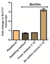
Mycobacterium tuberculosis biofilm upregulates Rv1717 expression upon sensing a dispersal signal. Transcripts of Rv1717 were estimated in a late log phase planktonic culture and biofilms of Mtb H37Rv wild type by quantitative RT-PCR. In case of biofilms, RNA was extracted before and after 1 h of replacing the used medium (depleting medium) with fresh medium with or without carbon source. Graph shows the linear fold change of Rv1717 transcripts calculated by the 2^▵▵Ct method using SigA transcripts for normalization. The values plotted are the mean ± SD of three biological replicates.

Similar articles

Cited by

References

    1. Arora K., Whiteford D. C., Lau-Bonilla D., Davitt C. M., Dahl J. L. (2008). Inactivation of lsr2 results in a hypermotile phenotype in Mycobacterium smegmatis. J. Bacteriol. 190, 4291–4300. 10.1128/JB.00023-08, PMID: - DOI - PMC - PubMed
    1. Baker P., Hill P. J., Snarr B. D., Alnabelseya N., Pestrak M. J., Lee M. J., et al. . (2016). Exopolysaccharide biosynthetic glycoside hydrolases can be utilized to disrupt and prevent Pseudomonas aeruginosa biofilms. Sci. Adv. 2:e1501632. 10.1126/sciadv.1501632, PMID: - DOI - PMC - PubMed
    1. Bales P. M., Renke E. M., May S. L., Shen Y., Nelson D. C. (2013). Purification and characterization of biofilm-associated EPS exopolysaccharides from ESKAPE organisms and other pathogens. PLoS One 8:e67950. 10.1371/journal.pone.0067950, PMID: - DOI - PMC - PubMed
    1. Basaraba R. J., Ojha A. K. (2017). Mycobacterial biofilms: revisiting tuberculosis bacilli in extracellular necrotizing lesions. Microbiol. Spectr. 5. 10.1128/microbiolspec.TBTB2-0024-2016, PMID: - DOI - PMC - PubMed
    1. Boles B. R., Horswill A. R. (2008). Agr-mediated dispersal of Staphylococcus aureus biofilms. PLoS Pathog. 4:e1000052. 10.1371/journal.ppat.1000052, PMID: - DOI - PMC - PubMed

LinkOut - more resources