. 2021 Jan 22:11:602530.
doi: 10.3389/fimmu.2020.602530. eCollection 2020.

Irradiated Tumor Fibroblasts Avoid Immune Recognition and Retain Immunosuppressive Functions Over Natural Killer Cells

Affiliations

Irradiated Tumor Fibroblasts Avoid Immune Recognition and Retain Immunosuppressive Functions Over Natural Killer Cells

Nannan Yang et al. Front Immunol. .

Abstract

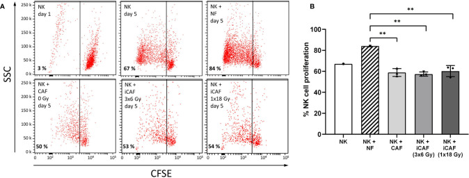
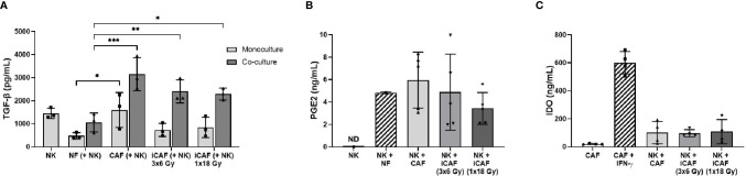
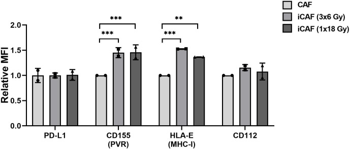
Recent studies have demonstrated that radiotherapy is able to induce anti-tumor immune responses in addition to mediating direct cytotoxic effects. Cancer-associated fibroblasts (CAFs) are central constituents of the tumor stroma and participate actively in tumor immunoregulation. However, the capacity of CAFs to influence immune responses in the context of radiotherapy is still poorly understood. This study was undertaken to determine whether ionizing radiation alters the CAF-mediated immunoregulatory effects on natural killer (NK) cells. CAFs were isolated from freshly resected non-small cell lung cancer tissues, while NK cells were prepared from peripheral blood of healthy donors. Functional assays to study NK cell immune activation included proliferation rates, expression of cell surface markers, secretion of immunomodulators, cytotoxic assays, as well as production of intracellular activation markers such as perforin and granzyme B. Our data show that CAFs inhibit NK cell activation by reducing their proliferation rates, the cytotoxic capacity, the extent of degranulation, and the surface expression of stimulatory receptors, while concomitantly enhancing surface expression of inhibitory receptors. Radiation delivered as single high-dose or in fractioned regimens did not reverse the immunosuppressive features exerted by CAFs over NK cells in vitro, despite triggering enhanced surface expression of several checkpoint ligands on irradiated CAFs. In summary, CAFs mediate noticeable immune inhibitory effects on cytokine-activated NK cells during co-culture in a donor-independent manner. However, ionizing radiation does not interfere with the CAF-mediated immunosuppressive effects.

Keywords: TME; cancer-associated fibroblasts; immune evasion; immunosuppression; immunotherapy; ionizing radiation; radiotherapy; tumor microenvironment.


Conflict of interest statement

The authors declare that the research was conducted in the absence of any commercial or financial relationships that could be construed as a potential conflict of interest.

Figures

Figure 1
Figure 1
Natural killer (NK) cell purity, cancer-associated fibroblast (CAF) characterization, and radiation-induced morphological changes in CAFs. (A) Dot plots from flow cytometer analyses, illustrating 93% purity of NK cells defined as CD56+CD3 upon negative selection by antibody-coated microbeads. (B) Immunostaining of cultivated CAFs, using CAF-specific FAP-1 antibody (red) and nuclear DAPI (blue). (C) Flow cytometry analyses of CAFs after immunostaining with CAF-specific αSMA antibody. Negative control is CAFs stained with isotype control antibody. (D) Representative culture of spindle-shaped, non-irradiated human lung CAFs at passage three. (E) Representative image of CAFs acquired 5 days after exposure to ionizing radiation (1x18 Gy). (F) Image of irradiated CAFs in culture, acquired 2 days after third dose of 6 Gy (3x6 Gy). (G) Culture of normal skin fibroblasts (NFs). (H) Image of non-adherent (CD56+CD3) NK cells in monoculture. (I) Image of co-cultures consisting of adherent CAFs and non-adherent NK cells.
Figure 2
Figure 2
Radiation-induced Fas-receptor on cancer-associated fibroblasts (CAFs) and natural killer (NK) cell-mediated killing. Panels in (A) show flow cytometry histograms comparing surface expression of Fas receptor in irradiated and non-irradiated CAFs from a representative donor. Control histograms represent CAFs exposed to isotype control antibodies. (B) Quantitative expression of Fas receptor presented as median fluorescence intensity (MFI) calculated from three independent CAF donors. (C) NK cell cytotoxicity assay measuring NK cell mediated cell killing of irradiated or non-irradiated CAFs as targets, in addition to two different control target cell types at NK to target ratio 5:1. Bars represent mean (± SD) from three independent CAF donors. Statistics and P-values between NF and CAFs were determined using one-way ANOVA with Tukey correction for multiple comparisons. ** indicates P < 0.01.
Figure 3
Figure 3
Effects of cancer-associated fibroblasts (CAFs) on natural killer (NK) cell proliferation. (A) Representative flow cytometry dot plots of the different experimental groups, showing percentage of carboxy-fluorescein succinimidyl ester (CSFE)-labeled NK cells on a side-scatter axis. (B) Bars representing mean (± SD) values for NK cell proliferation obtained from experiments with three different CAF donors. NK cells/NFs co-cultures were used as positive control, and the three experimental groups of NK cells/CAFs co-cultures were compared with the normal fibroblast (NF) group. Statistical P-values between NFs and CAFs co-cultures were determined using one-way ANOVA with Tukey correction for multiple comparisons. ** indicates P < 0.01.
Figure 4
Figure 4
Effects of cancer-associated fibroblasts (CAFs) on natural killer (NK) cell cytotoxic activity. Cytotoxic activity of NK cells co-cultured with fibroblasts was analyzed against CSFE-labeled K562 leukemic tumor cells at NK to target ratio of 5:1. Results were evaluated by flow cytometry and presented as percentage propidium-iodide positive (dead) cells versus side scatter activity (SSC). TGF-β treated NK cells served as control for repressed cytotoxic activity. Panels in (A) represent flow cytometry dot-plots from a representative experiment with one of the CAF donors. In (B), NK cell cytotoxic activity is represented as mean (± SD) values from flow experiments with three different CAF donors. Statistical P-values between mixed cultures with NF and CAFs were determined using one-way ANOVA with Tukey correction for multiple comparisons. Similarly, in (C), NK cell degranulation values are calculated as levels of LAMP-1 (CD107a) present on the NK cell surface after being employed against K562 leukemic tumor cells (for 4 h), at a NK to target ratio of 5:1, and an initial co-culturing with CAFs. Data represent mean (± SD) values from three different donors. * indicates P < 0.01.
Figure 5
Figure 5
Effects of cancer-associated fibroblasts (CAFs) on natural killer (NK) cell activation markers. NK cells were incubated in mixed cultures with NFs or CAFs. NK cells treated with TGF-β served as control of repressed functional activity. Following co-culturing, NK cells were analyzed for (A) intracellular expression of cytotoxic enzymes perforin and granzyme B after being employed against K562 cells (NK to K562 ratio 5:1); (B) intracellular expression of pro-inflammatory cytokines IFN-γ and TNF-α after exposure to K562 cells (5:1); and (C) released levels of IFN-γ and TNF-α in culture media (fibroblast to NK ratio 1:2). Results in (A, B) were measured by flow cytometry, whereas cytokine release in (C) was quantified by ELISA. For intracellular markers, signal intensities are expressed as median fluorescence intensity (MFI). Data represents mean (± SD) values from three different CAF donors. Statistical P-values between NF and CAFs were determined using one-way ANOVA with Tukey correction for multiple comparisons. Extracellular levels in (C) are presented as mean (± SD) values from four random CAF donors.
Figure 6
Figure 6
Effects of radiation on cancer-associated fibroblast (CAF)-derived immunomodulators. (A, B) Protein levels of secreted TGF-β and PGE2 in culture supernatants measured by ELISA. (C) Intracellular IDO protein expression in CAF cell lysates analyzed by ELISA. In (C), positive control is non-irradiated CAFs stimulated with IFN-γ and negative control non-irradiated CAFs cultured without stimulation. Data represents mean (± SD) values from three (A), five (B) or four (C) different CAF donors. P-values between NFs and CAFs in (A, B) were determined using one-way ANOVA with Tukey correction for multiple comparisons, whereas P-values between groups of monocultures and co-cultures in (C) were determined using two-way ANOVA with Dunnett correction for multiple comparisons. *P < 0.05, **P < 0.01 and ***P < 0.001.
Figure 7
Figure 7
Effects of cancer-associated fibroblasts (CAFs) on natural killer (NK) cell surface receptor expression. Following co-culturing with fibroblasts, isolated NK cells were analyzed by flow cytometry for their expression of NK cell surface markers (A) NKG2D, (B) NKp46, (C) DNAM-1, (D) NKG2A, (E) KIR2DL1 and (F) KIR3DL1. Results are presented as median fluorescence intensity (MFI) and data represents mean (± SD) values from experiments with three different CAF donors. Statistical P-values between NK/NFs and NK/CAF-groups were determined using one-way ANOVA with Tukey correction for multiple comparisons * and ** indicates P < 0.05 and P < 0.01, respectively.
Figure 8
Figure 8
Effects of radiation on cancer-associated fibroblast (CAF) surface expression of checkpoint molecules. Surface expression of checkpoint ligands PD-L1, CD155, CD112, and HLA-E (MHC-I) were analyzed by flow cytometry on control and irradiated CAFs (iCAFs). Results are expressed as fold non-irradiated CAF median fluorescence intensity (MFI). Bars represent mean (± SD) values from two different CAF donors. Statistical P-values between control CAFs and irradiated CAF-groups were determined using two-way ANOVA with Dunnett correction for multiple comparisons. **P < 0.01 and ***P < 0.001.

References

    1. Tree AC, Khoo VS, Eeles RA, Ahmed M, Dearnaley DP, Hawkins MA, et al. Stereotactic body radiotherapy for oligometastases. Lancet Oncol (2013) 14(1):e28–37. 10.1016/S1470-2045(12)70510-7 - DOI - PubMed
    1. Weichselbaum RR, Liang H, Deng L, Fu Y-X. Radiotherapy and immunotherapy: a beneficial liaison? Nat Rev Clin Oncol (2017) 14(6):365–79. 10.1038/nrclinonc.2016.211 - DOI - PubMed
    1. Pitroda SP, Chmura SJ, Weichselbaum RR. Integration of radiotherapy and immunotherapy for treatment of oligometastases. Lancet Oncol (2019) 20(8):e434–42. 10.1016/S1470-2045(19)30157-3 - DOI - PubMed
    1. Ko EC, Raben D, Formenti SC. The Integration of Radiotherapy with Immunotherapy for the Treatment of Non-Small Cell Lung Cancer. Clin Cancer Res (2018) 24(23):5792–806. 10.1158/1078-0432.CCR-17-3620 - DOI - PubMed
    1. Bang A, Schoenfeld JD, Sun AY. PACIFIC: shifting tides in the treatment of locally advanced non-small cell lung cancer. Transl Lung Cancer Res (2019) 8(Suppl 2):S139–46. 10.21037/tlcr.2019.09.04 - DOI - PMC - PubMed

Publication types

MeSH terms