Review
. 2021 Jan 28:11:613815.
doi: 10.3389/fimmu.2020.613815. eCollection 2020.

Mechanisms Governing Immunotherapy Resistance in Pancreatic Ductal Adenocarcinoma

Affiliations
Review

Mechanisms Governing Immunotherapy Resistance in Pancreatic Ductal Adenocarcinoma

Zoe C Schmiechen et al. Front Immunol. .

Abstract

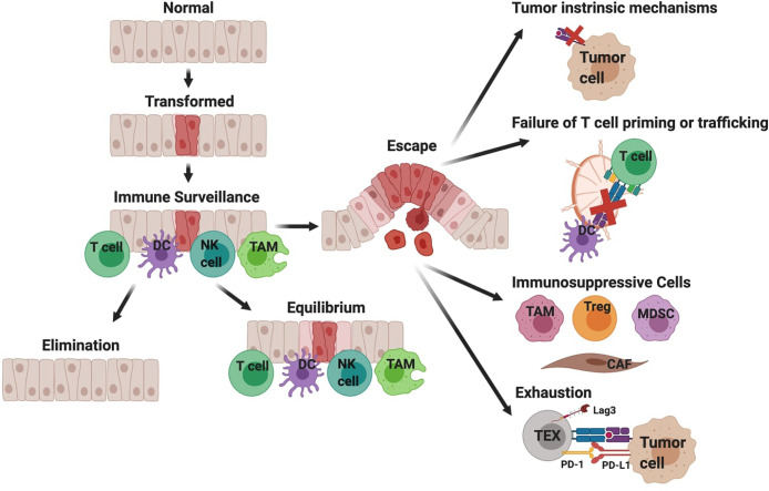
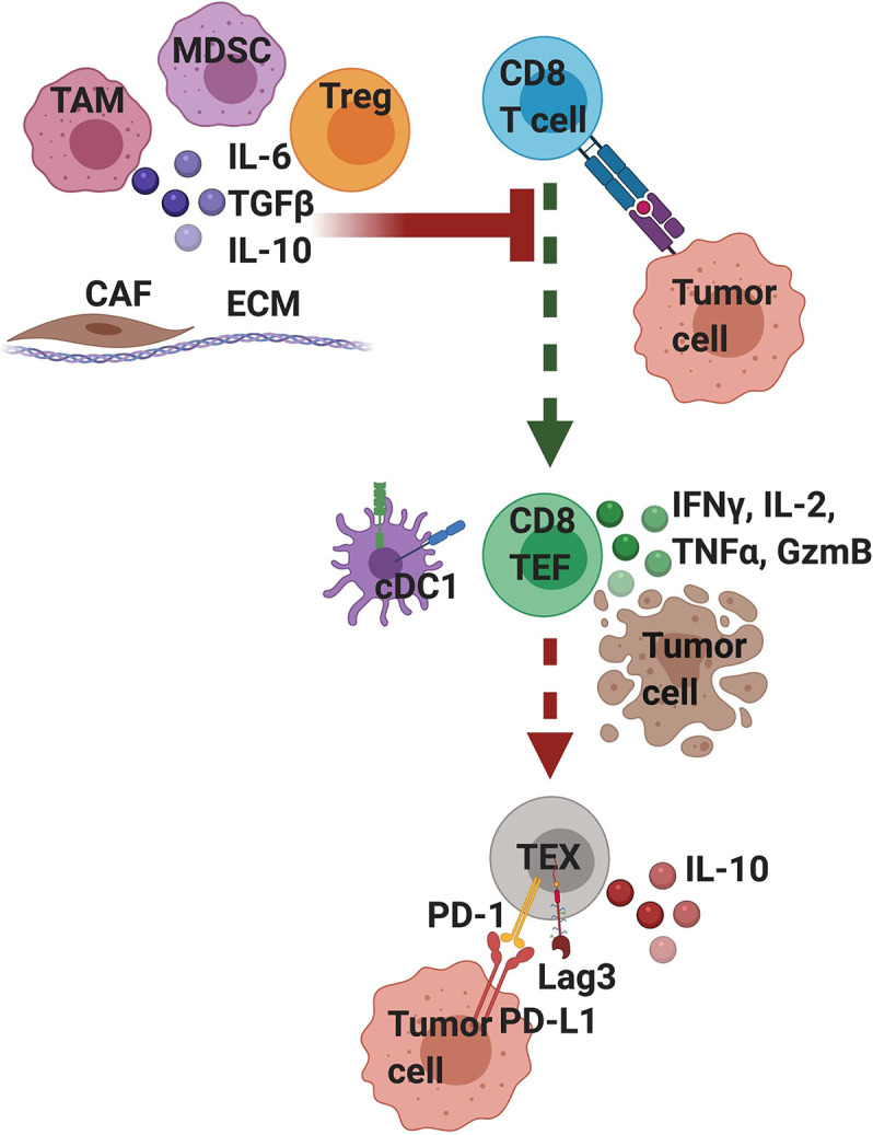
Pancreatic ductal adenocarcinoma (PDA) is a lethal malignancy with an overall 5-year survival rate of 10%. Disease lethality is due to late diagnosis, early metastasis and resistance to therapy, including immunotherapy. PDA creates a robust fibroinflammatory tumor microenvironment that contributes to immunotherapy resistance. While previously considered an immune privileged site, evidence demonstrates that in some cases tumor antigen-specific T cells infiltrate and preferentially accumulate in PDA and are central to tumor cell clearance and long-term remission. Nonetheless, PDA can rapidly evade an adaptive immune response using a myriad of mechanisms. Mounting evidence indicates PDA interferes with T cell differentiation into potent cytolytic effector T cells via deficiencies in naive T cell priming, inducing T cell suppression or promoting T cell exhaustion. Mechanistic research indicates that immunotherapy combinations that change the suppressive tumor microenvironment while engaging antigen-specific T cells is required for treatment of advanced disease. This review focuses on recent advances in understanding mechanisms limiting T cell function and current strategies to overcome immunotherapy resistance in PDA.

Keywords: PD-1; PD-L1; T cell; exhaustion; immunosuppression; immunotherapy; pancreatic cancer; pancreatic ductal adenocarcinoma.


Conflict of interest statement

IS serves on the scientific advisory board for Luminary Therapeutics and Immunogenesis. The remaining author declares that the research was conducted in the absence of any commercial or financial relationships that could be construed as a potential conflict of interest.

Figures

Figure 1
Figure 1
Simplified overview of immune surveillance and tumor evasion in pancreatic ductal adenocarcinoma (PDA). Immune surveillance is the process whereby the immune system surveys the body for malignant or infected cells. Components of immune surveillance include T cells, DCs, NK cells, and macrophages (TAM). Mutations in oncogene KRAS are a driver of PDA. The immune response to transformed tissue can either result in complete elimination of the malignancy, equilibrium to prevent further growth of the malignancy, or escape and development of clinically significant tumors. This figure provides a hypothesized sequence of immune evasion, but this process is likely not linear and instead a dynamic progression. Cancer cells can escape T cell recognition by losing target antigen expression and/or developing defects in antigen processing and presentation. Additionally, defects in T cell priming or trafficking to tumors may limit antigen-specific T cell responses and can be attributed to insufficient mature DCs. When antigen-specific T cells successfully infiltrate tumors, their function may be limited by immunosuppressive cytokines produced by macrophages (TAM, tumor-associated macrophage), regulatory T cells (Treg), myeloid-derived suppressor cells (MDSC), or cancer associated fibroblasts (CAF). Lastly chronic T cell receptor signaling drives T cell exhaustion, resulting in reduced effector function.
Figure 2
Figure 2
T cell intrinsic and extrinsic factors influence T cell differentiation and functionality in pancreatic cancer. Tumor-infiltrating T cells that strongly recognize tumor antigen mediate transient anti-tumor activity but if the tumor is not cleared, differentiate into exhausted (TEX) T cells, which is driven by persistent T cell receptor (TCR) signaling. Exhausted T cells are often characterized by their expression of PD-1 and Lag3 and are hypofunctional. Tumor cells and other stromal cells can express ligands for these inhibitory receptors, such as PD-L1, a ligand for PD- 1. Signaling through these inhibitory receptors interferes with T cell function and differentiation state. Moreover, exhausted T cells may participate in their own suppression by producing IL-10. A variety of extrinsic cells and factors enriched in the suppressive tumor microenvironment can interfere with TCR signaling and activation. These include tumor associated macrophages (TAM), myeloid-derived suppressor cells (MDSC), regulatory T cells (Tregs), and cancer associated fibroblasts (CAF) all embedded within a dense extracellular matrix (ECM). Immunosuppressive cells secrete cytokines including IL-10 and TGFβ that can suppress T cell activation and TCR signaling. Mechanistically, extrinsic suppression and TCR-driven exhaustion are distinct processes that converge to suppress tumor immunity. We posit that targeting immunosuppression may lead to only transient anti-tumor immune responses because if the tumor is not cleared; T cells will become exhausted. Thus, PDA likely requires combination immunotherapies that target both T cell extrinsic immunosuppression and T cell intrinsic exhaustion.
Figure 3
Figure 3
Mechanisms of immune escape in pancreatic cancer. Simplified overview of mechanisms by which pancreatic ductal adenocarcinoma (PDA) evades the immune response and examples of potential therapeutic strategies to target these mechanisms. Some strategies may obviate multiple tumor evasion mechanisms. Combination strategies may be necessary to promote synergy and overcome tumor evasion from the immune system.

Similar articles

Cited by

References

    1. Howlader N, Noone A, Krapcho M, Miller D, Brest A, Yu M, et al. SEER Cancer Statistics Review, 1975-2016. Bethesda, MD: National Cancer Institute; (2019). Available at: https://seer.cancer.gov/csr/1975_2016/.
    1. Stromnes IM, DelGiorno KE, Greenberg PD, Hingorani SR. Stromal reengineering to treat pancreas cancer. Carcinogenesis (2014) 35:1451–60. 10.1093/carcin/bgu115 - DOI - PMC - PubMed
    1. Ryan DP, Hong TS, Bardeesy N. Pancreatic adenocarcinoma. N Engl J Med (2014) 371:1039–49. 10.1056/NEJMra1404198 - DOI - PubMed
    1. Huang L, Jansen L, Balavarca Y, van der Geest L, Lemmens L, Van Eycken L, et al. Stratified survival of resected and overall pancreatic cancer patients in Europe and the USA in the early twenty-first century: a large, international population-based study Lei. BMC Med (2018) 16. 10.1186/s12916-018-1120-9 - DOI - PMC - PubMed
    1. Rahib L, Smith BD, Aizenberg R, Rosenzweig AB, Fleshman JM, Matrisian LM, et al. Projecting cancer incidence and deaths to 2030: The unexpected burden of thyroid, liver, and pancreas cancers in the united states. Cancer Res (2014) 74:2913–21. 10.1158/0008-5472.CAN-14-0155 - DOI - PubMed

Publication types

MeSH terms