

Cytokine Signature Induced by SARS-CoV-2 Spike Protein in a Mouse Model
- PMID: 33584719
- PMCID: PMC7876321
- DOI: 10.3389/fimmu.2020.621441
Cytokine Signature Induced by SARS-CoV-2 Spike Protein in a Mouse Model
Abstract
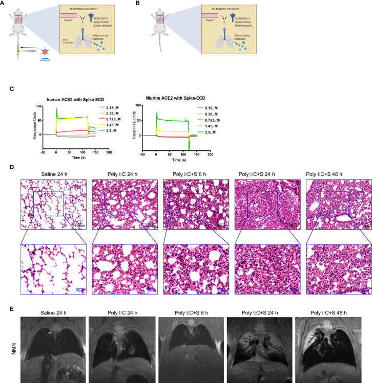
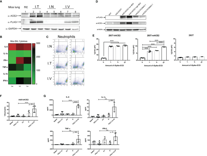
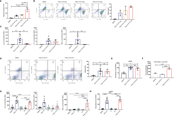
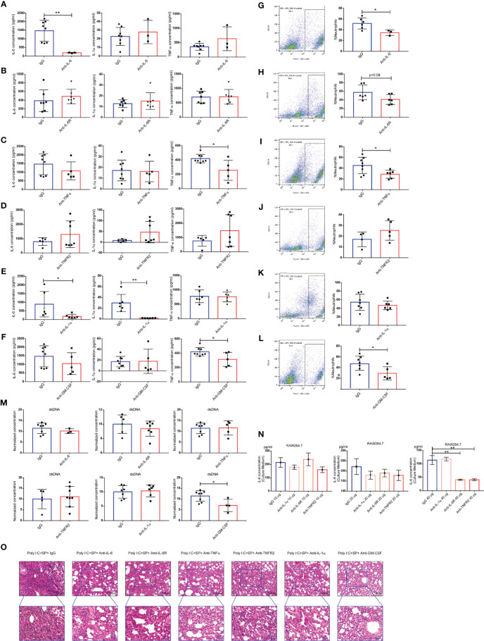
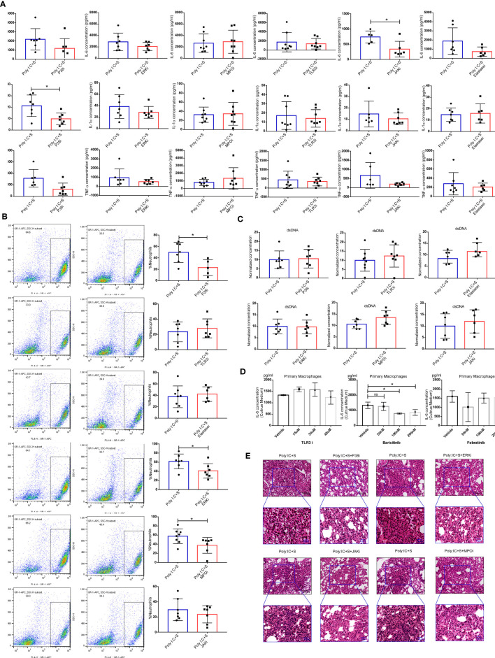
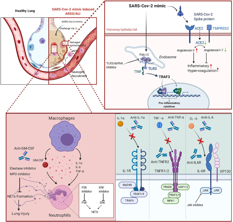
Although COVID-19 has become a major challenge to global health, there are currently no efficacious agents for effective treatment. Cytokine storm syndrome (CSS) can lead to acute respiratory distress syndrome (ARDS), which contributes to most COVID-19 mortalities. Research points to interleukin 6 (IL-6) as a crucial signature of the cytokine storm, and the clinical use of the IL-6 inhibitor tocilizumab shows potential for treatment of COVID-19 patient. In this study, we challenged wild-type and adenovirus-5/human angiotensin-converting enzyme 2-expressing BALB/c mice with a combination of polyinosinic-polycytidylic acid and recombinant SARS-CoV-2 spike-extracellular domain protein. High levels of TNF-α and nearly 100 times increased IL-6 were detected at 6 h, but disappeared by 24 h in bronchoalveolar lavage fluid (BALF) following immunostimulant challenge. Lung injury observed by histopathologic changes and magnetic resonance imaging at 24 h indicated that increased TNF-α and IL-6 may initiate CSS in the lung, resulting in the continual production of inflammatory cytokines. We hypothesize that TNF-α and IL-6 may contribute to the occurrence of CSS in COVID-19. We also investigated multiple monoclonal antibodies (mAbs) and inhibitors for neutralizing the pro-inflammatory phenotype of COVID-19: mAbs against IL-1α, IL-6, TNF-α, and granulocyte-macrophage colony-stimulating factor (GM-CSF), and inhibitors of p38 and JAK partially relieved CSS; mAbs against IL-6, TNF-α, and GM-CSF, and inhibitors of p38, extracellular signal-regulated kinase, and myeloperoxidase somewhat reduced neutrophilic alveolitis in the lung. This novel murine model opens a biologically safe, time-saving avenue for clarifying the mechanism of CSS/ARDS in COVID-19 and developing new therapeutic drugs.
Keywords: COVID-19; SARS-CoV-2; acute respiratory distress syndrome; cytokine storm syndrome; murine model.
Copyright © 2021 Gu, Zhao, Jin, Song, Zhi, Zhao, Ma, Zheng, Wang, Liu, Xin, Han, Li, Dong, Liu and Dong.
Conflict of interest statement
The authors declare that the research was conducted in the absence of any commercial or financial relationships that could be construed as a potential conflict of interest.
Figures
Similar articles
-
Two new and effective food-extracted immunomodulatory agents exhibit anti-inflammatory response activity in the hACE2 acute lung injury murine model of COVID-19.Front Immunol. 2024 May 14;15:1374541. doi: 10.3389/fimmu.2024.1374541. eCollection 2024. Front Immunol. 2024. PMID: 38807598 Free PMC article.
-
Induction of Exaggerated Cytokine Production in Human Peripheral Blood Mononuclear Cells by a Recombinant SARS-CoV-2 Spike Glycoprotein S1 and Its Inhibition by Dexamethasone.Inflammation. 2021 Oct;44(5):1865-1877. doi: 10.1007/s10753-021-01464-5. Epub 2021 Apr 16. Inflammation. 2021. PMID: 33860869 Free PMC article.
-
SARS-CoV-2 nucleocapsid protein, rather than spike protein, triggers a cytokine storm originating from lung epithelial cells in patients with COVID-19.Infection. 2024 Jun;52(3):955-983. doi: 10.1007/s15010-023-02142-4. Epub 2023 Dec 22. Infection. 2024. PMID: 38133713 Free PMC article.
-
Critical Determinants of Cytokine Storm and Type I Interferon Response in COVID-19 Pathogenesis.Clin Microbiol Rev. 2021 May 12;34(3):e00299-20. doi: 10.1128/CMR.00299-20. Print 2021 Jun 16. Clin Microbiol Rev. 2021. PMID: 33980688 Free PMC article. Review.
-
The role of Interleukin 6 inhibitors in therapy of severe COVID-19.Biomed Pharmacother. 2020 Nov;131:110698. doi: 10.1016/j.biopha.2020.110698. Epub 2020 Aug 28. Biomed Pharmacother. 2020. PMID: 32920514 Free PMC article. Review.
Cited by
-
Dichotomous Role of Tumor Necrosis Factor in Pulmonary Barrier Function and Alveolar Fluid Clearance.Front Physiol. 2022 Feb 21;12:793251. doi: 10.3389/fphys.2021.793251. eCollection 2021. Front Physiol. 2022. PMID: 35264975 Free PMC article. Review.
-
Mechanistic Understanding of Lung Inflammation: Recent Advances and Emerging Techniques.J Inflamm Res. 2022 Jun 15;15:3501-3546. doi: 10.2147/JIR.S282695. eCollection 2022. J Inflamm Res. 2022. PMID: 35734098 Free PMC article. Review.
-
Human Lung Fibroblasts Exhibit Induced Inflammation Memory via Increased IL6 Gene Expression and Release.Front Immunol. 2022 Jul 22;13:921728. doi: 10.3389/fimmu.2022.921728. eCollection 2022. Front Immunol. 2022. PMID: 35941890 Free PMC article.
-
Two Years into the COVID-19 Pandemic: Lessons Learned.ACS Infect Dis. 2022 Sep 9;8(9):1758-1814. doi: 10.1021/acsinfecdis.2c00204. Epub 2022 Aug 8. ACS Infect Dis. 2022. PMID: 35940589 Free PMC article. Review.
-
Exposure to COVID-19 virus-like particles modulates firing patterns of cortical neurons in the mouse brain.Commun Biol. 2025 Jul 3;8(1):993. doi: 10.1038/s42003-025-08435-8. Commun Biol. 2025. PMID: 40610705 Free PMC article.
References
-
- Callejas Rubio JL, Luna Del Castillo JD, de la Hera Fernandez J, Guirao Arrabal E, Colmenero Ruiz M, Ortego Centeno N. Effectiveness of corticoid pulses in patients with cytokine storm syndrome induced by SARS-CoV-2 infection. Med Clin (2020) 155(4):159–61. 10.1016/j.medcle.2020.07.002 - DOI - PMC - PubMed
-
- Bhargava P, Panda P, Ostwal V, Ramaswamy A. Repurposing valproate to prevent acute respiratory distress syndrome/acute lung injury in COVID-19: A review of immunomodulatory action. Cancer Res Statistics Treat (2020) 3(5):65. 10.4103/CRST.CRST_156_20 - DOI
MeSH terms
Substances
LinkOut - more resources
Full Text Sources
Other Literature Sources
Medical
Research Materials
Miscellaneous