Review
. 2021 Jan 27:11:621931.
doi: 10.3389/fimmu.2020.621931. eCollection 2020.

Interleukin-1 as Innate Mediator of T Cell Immunity

Affiliations
Review

Interleukin-1 as Innate Mediator of T Cell Immunity

Bram Van Den Eeckhout et al. Front Immunol. .

Abstract

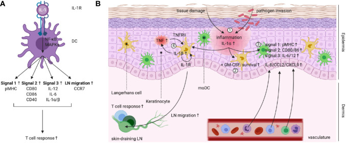
The three-signal paradigm tries to capture how the innate immune system instructs adaptive immune responses in three well-defined actions: (1) presentation of antigenic peptides in the context of MHC molecules, which allows for a specific T cell response; (2) T cell co-stimulation, which breaks T cell tolerance; and (3) secretion of polarizing cytokines in the priming environment, thereby specializing T cell immunity. The three-signal model provides an empirical framework for innate instruction of adaptive immunity, but mainly discusses STAT-dependent cytokines in T cell activation and differentiation, while the multi-faceted roles of type I IFNs and IL-1 cytokine superfamily members are often neglected. IL-1α and IL-1β are pro-inflammatory cytokines, produced following damage to the host (release of DAMPs) or upon innate recognition of PAMPs. IL-1 activity on both DCs and T cells can further shape the adaptive immune response with variable outcomes. IL-1 signaling in DCs promotes their ability to induce T cell activation, but also direct action of IL-1 on both CD4+ and CD8+ T cells, either alone or in synergy with prototypical polarizing cytokines, influences T cell differentiation under different conditions. The activities of IL-1 form a direct bridge between innate and adaptive immunity and could therefore be clinically translatable in the context of prophylactic and therapeutic strategies to empower the formation of T cell immunity. Understanding the modalities of IL-1 activity during T cell activation thus could hold major implications for rational development of the next generation of vaccine adjuvants.

Keywords: CD4+ T cells; CD8+ T cells; cancer immunotherapy; cellular adjuvant; dendritic cells; interleukin-1; vaccination.


Conflict of interest statement

JT is affiliated with Orionis Biosciences BV as scientific advisor and holds equity interests in Orionis Biosciences BV. The remaining authors declare that the research was conducted in the absence of any commercial or financial relationships that could be construed as a potential conflict of interest.

Figures

Figure 1
Figure 1
Synthesis, modification and extracellular release of human (pro-)IL-1α. (1) and (2) Inflammatory triggers can induce pro-IL-1α expression, whereas homeostatic expression is regulated by housekeeping transcription factors, such as Sp1. (3) Alternatively, expression can be initiated following DNA release of negative regulators. (4) Pro-IL-1α is modified post-translationally. (5) Pro-IL-1α continuously shuttles between cytosol and nucleus and nuclear translocation is mediated via HAX-1, which binds to the pro-IL-1α NLS. Binding of pro-IL-1α to chromatin sequesters the cytokine in the nucleus, whereas interaction with histone modifying enzymes mediates pro-inflammatory gene expression. (6) Calpain cleaves pro-IL-1α in the cytosol, leading to the formation of mature and fully biologically active IL-1α. (7) and (8) Following accidental cell death or damage, (pro-)IL-1α is released extracellularly, where maturation by cleavage is mediated by granzyme B, neutrophil elastases and mast cell chymases. (9) Post-translational modification can allow for extracellular membrane anchorage of pro-IL-1α. (10) A unique intracellularly occurring form of IL-1R2 can neutralize and sequester pro-IL-1α in the cytosol, possibly by masking its NLS. (11) Turnover of (pro-)IL-1α and the cleaved N-terminal pro-piece is mediated in the proteasome. Figure adapted from (35). Created with BioRender.com.
Figure 2
Figure 2
The two-step paradigm of controlled IL-1β release. (1) Signal 1 or “priming” involves expression of pro-IL-1β, NLRP3 and caspase 1 following triggering of pro-inflammatory receptor complexes (e.g. TLR4 or IL-1R) and activation of the transcription factor NF-κB. (2) NLRP3’s LRR falls back onto the NACHT domain, rendering the protein inactive in the cytoplasm. (3) Signal 2 or “activation” can be mediated by PAMPs (e.g. viral RNA) or DAMPs (e.g. extracellular ATP, extracellular particles and crystal complexes). Viral RNA can activate mitochondrial antiviral signaling (MAVS) protein. ATP is detected via the P2RX7 receptor and extracellular particles and crystal complexes can induce intracellular lysosome disruption, leading to changes in Ca2+ flux and K+ efflux. Intracellular changes in ion concentration can influence mitochondria, as such inducing release of ROS and oxidized mtDNA or relocalization of cardiolipin to the outer mitochondrial membrane. (4) and (5) Relocalized cardiolipin, oxidized mDNA, activated MAVS protein and changes in cytosolic ion concentrations can induce conformational changes in NLRP3 and lead to formation of the activated NLRP3 inflammasome after recruitment of NEK7, ASC and caspase 1. (6) Activated caspase 1 cleaves immature pro-IL-1β to mature and fully biologically active IL-1β. In addition, caspase 1 can cleave the GSDMD protein. (7) GSDMDNterm integrates in the membrane as a pore, which induces pyroptosis and represents a putative way for IL-1β release. CD, catalytic domain; NTE, N-terminal extension. Figure adapted from (50). Created with BioRender.com.
Figure 3
Figure 3
Formation of the signaling-competent IL-1R complex and downstream IL-1 signaling. (1) Extracellular IL-1β makes two contacts with IL-1R1 and induces a conformational change in the primary receptor. (2) This allows IL-1R1 to recruit and bind to co-receptor IL-1R3. (3) Receptor dimerization leads to scaffolding of intracellular receptor TIR domains via the R-interface. (4) and (5) Formation of an intracellular TIR scaffold allows for recruitment of the MyD88 adaptor molecule via S-interface interactions. MyD88 comprises an N-terminal death domain (DD), an intermediate domain (ID) and a C-terminal TIR domain. IRAK-1, IRAK-4 and/or IRAK-2 are recruited. These molecules comprise an N-terminal DD and a C-terminal catalytic domain. Under steady-state, IRAK-1 is bound to Tollip. As such, the Myddosome is formed. (6) IRAK-4 auto-phosphorylates and gains full activity, in turn activating IRAK-1 by phosphorylation. This induces rapid IRAK-1 auto-phosphorylation. (7) A conformational change in IRAK-1 allows its dissociation from Tollip. Different IRAK-1 oligomers form a scaffold for TRAF6, which becomes activated upon oligomerization. UBC13 and UEV1A facilitate TRAF6 self-poly-ubiquitination (K63-linked). TAB2 and TAB3 recruit TAB1 and the MAP3K TAK1 to the K63-linked poly-ubiquitin chain on TRAF6. (8) TRAF6 poly-ubiquitinates TAK1. (9) Initiation of the canonical NF-κB pathway: NEMO binds to the poly-ubiquitin chain on TAK1 and recruits IKKα and IKKβ. IKKα and IKKβ become activated following phosphorylation by TAK1. (10) and (11) IKKα and IKKβ phosphorylate IκBα, which allows for its dissociation from p50/p65 NF-κB. (12) Free p50/p65 NF-κB translocates to the nucleus and binds to specific κB sites on DNA, as such inducing gene expression. (13) Initiation of the MAPK pathway: the MAP3K MAP3K3 binds the poly-ubiquitin chain on TAK1. MAP3K3 becomes activated following phosphorylation by TRAF6. IKKα and IKKβ activate TPL2 by phosphorylation. MAP3K3, TAK1 and TPL2 activate different MAP2Ks by phosphorylation. (14) MAP2Ks activate the p38, JNK and ERK MAPKs by phosphorylation. (15) In turn, MAPKs activate different transcription factors by phosphorylation, as such inducing gene expression. (16) Binding of IL-1β to sIL-1R2 does not induce signal transduction. (17) Binding of IL-1β to the signaling-incompetent IL-1R complex does not induce signal transduction. (18) Binding of IL-1Ra to the signaling-competent IL-1R complex does not induce signal transduction. Created with BioRender.com.
Figure 4
Figure 4
(A) Left scheme: IL-1R triggering in DCs empowers their capacity to promote T cell responses. (B) IL-1 activity plays a central role during the keratinocyte-LC cross-talk in the skin. (1) Damage to keratinocytes following tissue damage or pathogen invasion leads to the release of (pro-)IL-1α. (2) In synergy with tissue-derived GM-CSF, IL-1R signaling promotes LC survival. (3) IL-1R signaling in moDCs upregulates pMHC (signal 1), expression of co-stimulatory molecules (signal 2), production of priming cytokines (signal 3) and release of chemokines, leading to attraction of neutrophils, monocytes and lymphocytes. (4) PRR and IL-1R signaling promotes LC IL-1β production. IL-1β acts on keratinocytes and induces TNF release, which in turns signals via TNFRII on LCs. TNFRII and autocrine IL-1R signaling in LCs enable migration to the skin-draining LNs, where T cell responses can be initiated. Created with BioRender.com.
Figure 5
Figure 5
IL-1 signaling facilitates TH17 differentiation. TGF-β drives the expression of both FoxP3 and RORγT during CD4+ T cell priming. Commitment to a role as regulator or effector is further dependent on IL-2 (STAT5) and IL-6 (STAT3) activity, which antagonize each other. This process was found to be facilitated by other environmental factors, including the metabolite retinoic acid (RA) and the pro-inflammatory cytokine IL-1. Priming environments enriched in TGF-β concentrations and lacking STAT3-dependent cytokines drive Treg development. IL-2 activates STAT5, leading to sustained FoxP3 expression and inhibition of IL-17A and IL-17F production. Presence of RA empowers IL-2 expression and further pushes the balance towards the Treg phenotype. Priming environments that include IL-6 drive STAT3 activation, which mediates IL-17A and IL-17F expression and inhibits FoxP3 expression. IL-1 activates NF-κB, which inhibits expression of SOCS3. This further empowers STAT3 activity and pushes the balance towards the TH17 phenotype. In the absence of TGF-β, development of a TH1-like TH17 phenotype can be driven by IL-6, IL-23 (STAT3), and IL-1. This pathogenic phenotype is characterized by production of IL-17, IFN-γ, IL-21, and IL-22. Figure adapted from (176, 177). Created with BioRender.com.
Figure 6
Figure 6
The whereabouts of IL-1R signaling during stimulation of CD8+ T cell responses. While IL-1 activity can potently promote CD8+ T cell responses at multiple levels, the exact whereabouts of IL-1R triggering for mediation of these effects remain under debate. This figure indicates the context-dependency of several key findings: (A) CD8+ T cell responses during LCMV-Armstrong infection; (B) Treatment of naive mice with antigen and IL-1β following ATCT of CD8+ T cells (240); (C) Treatment of B16 melanoma tumor-bearing mice that received ATCT of CD8+ T cells with IL-1β (241); (D) CD8+ T cell responses during IAV infection (115). Created with BioRender.com.

References

    1. Iwasaki A, Medzhitov R. Control of adaptive immunity by the innate immune system. Nat Immunol (2015) 16:343–53. 10.1038/ni.3123 - DOI - PMC - PubMed
    1. Janeway CA. a. Pillars article: approaching the asymptote? Evolution and revolution in immunology. Cold spring harb symp quant biol. 1989. 54: 1-13. J Immunol (2013) 191:4475–87. - PubMed
    1. Matzinger P. Tolerance, danger and the extended family. Annu Rev Immunol (1994) 12:991–1045. 10.1146/annurev.iy.12.040194.005015 - DOI - PubMed
    1. Jain A, Pasare C. Innate control of adaptive immunity: Beyond the three-signal paradigm. J Immunol (2017) 198:3791–800. 10.4049/jimmunol.1602000 - DOI - PMC - PubMed
    1. Gilbert SC. T-cell-inducing vaccines - what’s the future. Immunology (2012) 135:19–26. 10.1111/j.1365-2567.2011.03517.x - DOI - PMC - PubMed

Publication types

MeSH terms