. 2021 Jan 28:11:622385.
doi: 10.3389/fimmu.2020.622385. eCollection 2020.

Self-Amplifying Pestivirus Replicon RNA Encoding Influenza Virus Nucleoprotein and Hemagglutinin Promote Humoral and Cellular Immune Responses in Pigs

Affiliations

Self-Amplifying Pestivirus Replicon RNA Encoding Influenza Virus Nucleoprotein and Hemagglutinin Promote Humoral and Cellular Immune Responses in Pigs

Thomas Démoulins et al. Front Immunol. .

Abstract

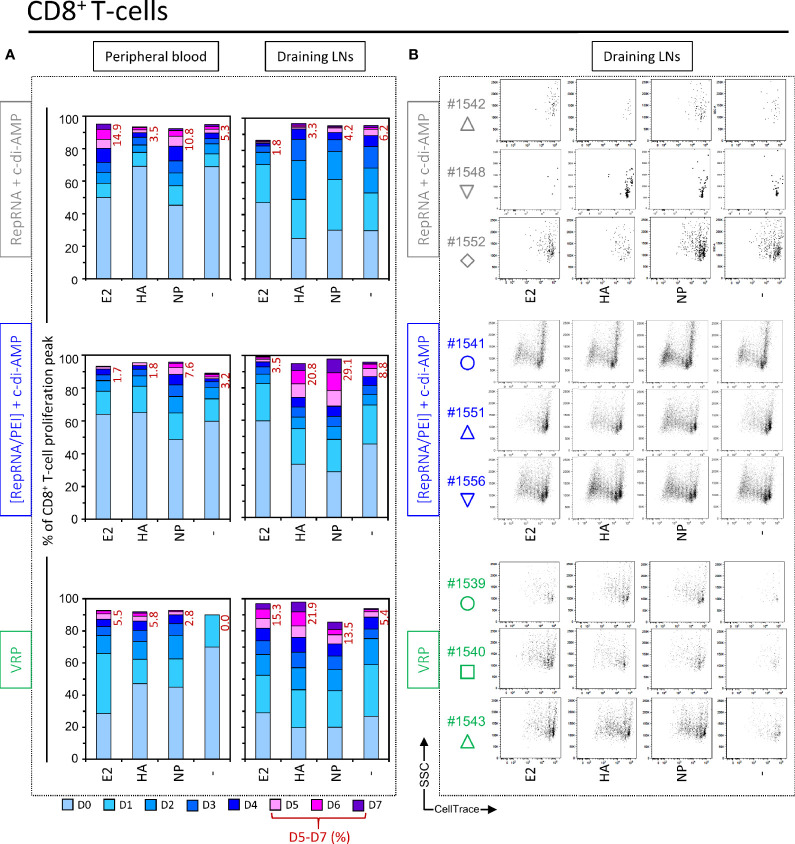
Self-amplifying replicon RNA (RepRNA) promotes expansion of mRNA templates encoding genes of interest through their replicative nature, thus providing increased antigen payloads. RepRNA derived from the non-cytopathogenic classical swine fever virus (CSFV) targets monocytes and dendritic cells (DCs), potentially promoting prolonged antigen expression in the DCs, contrasting with cytopathogenic RepRNA. We engineered pestivirus RepRNA constructs encoding influenza virus H5N1 (A/chicken/Yamaguchi/7/2004) nucleoprotein (Rep-NP) or hemagglutinin (Rep-HA). The inherent RNase-sensitivity of RepRNA had to be circumvented to ensure efficient delivery to DCs for intracellular release and RepRNA translation; we have reported how only particular synthetic delivery vehicle formulations are appropriate. The question remained concerning RepRNA packaged in virus replicon particles (VRPs); we have now compared an efficient polyethylenimine (PEI)-based formulation (polyplex) with VRP-delivery as well as naked RepRNA co-administered with the potent bis-(3',5')-cyclic dimeric adenosine monophosphate (c-di-AMP) adjuvant. All formulations contained a Rep-HA/Rep-NP mix, to assess the breadth of both humoral and cell-mediated defences against the influenza virus antigens. Assessment employed pigs for their close immunological relationship to humans, and as natural hosts for influenza virus. Animals receiving the VRPs, as well as PEI-delivered RepRNA, displayed strong humoral and cellular responses against both HA and NP, but with VRPs proving to be more efficacious. In contrast, naked RepRNA plus c-di-AMP could induce only low-level immune responses, in one out of five pigs. In conclusion, RepRNA encoding different influenza virus antigens are efficacious for inducing both humoral and cellular immune defences in pigs. Comparisons showed that packaging within VRP remains the most efficacious for delivery leading to induction of immune defences; however, this technology necessitates employment of expensive complementing cell cultures, and VRPs do not target human cells. Therefore, choosing the appropriate synthetic delivery vehicle still offers potential for rapid vaccine design, particularly in the context of the current coronavirus pandemic.

Keywords: c-di-AMP adjuvant; humoral and cellular immune response; influenza vaccines; polyplexes; self-amplifying replicon RNA; virus replicon particle.


Conflict of interest statement

Application of delivery vehicles for the delivery of RepRNA vaccines to DCs (23) using replicons derived from classical swine fever virus, as employed in this paper, has been filed for patents in Europe, USA, Canada, and India, with priority date of 2008. The filing was by the authors KM and NR, together with Jon Duri Tratschin (all three as inventors) (WO 2009146867) (6), and assigned to their employer – the Institute of Virology and Immunology. This does not alter the authors’ adherence to the policies of sharing data and materials. CG and TE are named as inventors in a patent application covering the use of c-di-AMP as adjuvant (PCT/EP 2006010693). The remaining authors declare that the research was conducted in the absence of any commercial or financial relationships that could be construed as a potential conflict of interest.

Figures

Figure 1
Figure 1
Prime-boost protocol for i.d. pig immunization. (A) Schematic representation of the CSFV genome (parent virus genome) employed for generating the RepRNA constructs: the deletion of the Erns coding sequence (ΔErns) and the inserted GOI (Rep-NP, Rep-HA) are shown. (B) Illustration of RepRNA functional assay by confocal microscopy, adapted from the infection center assay (ICA) reported elsewhere (25). In this example, SK-6 cells were electroporated with Rep-NP, after what we performed a tenfold serial dilution of the transfected cells (corresponding to 10-1 µg RepRNA and 8 x 10⁵ cells per 100 µl in the first dilution to 10-4 µg RepRNA and 800 cells per 100 µl in the 4th dilution). 100 µl of each dilution containing electroporated cells were transferred to the corresponding Lab-Tek® wells and let to adhere to 3 x 10⁵ pre-seeded SK-6 cells for 4–6 h in the 37°C incubator in MEM/glutamax/7%v/v-horse-serum. Then, the medium was replaced, and cells were cultured for 48 h at 37°C. The staining protocol involved fixation, permeabilisation and labelling with antibody against NP (green) and E2 (red); cell surfaces were stained with WGA-Alexa633 (gray). (C) Illustration of the prime-boost protocol for i.d. pig immunization. On day 56 the peripheral blood and the draining superficial cervical dorsal LNs were collected from sacrificed pigs.
Figure 2
Figure 2
Virus replicon particle (VRP) and NGA deliver RepRNA for translation in vivo. (A, B) Mice and rabbits were injected subcutaneously at 0, 14, and 28 days. Rep-HA and Rep-NP were delivered in NGA or VRP; all vaccines were adjuvanted with c-di-AMP adjuvant. Serum samples were assessed for anti-HA and anti-NP antibodies by ELISA and titers estimated at the times shown. (C) Adoptive transfer studies in mice have been performed in order to investigate the potential of the polyethylenimine (PEI) formulations to stimulate cellular immune responses. In brief, 24 h prior vaccination of wild type mice, CFSE labeled Thy1.1 CD8+ T cells of antigen-specific TCR transgenic mice were injected intravenously. 7 days later, cervical LNs of transplanted mice were collected and the proliferative capacity of the CFSE labeled Thy1.1 CD8+ T cells was analyzed by flow cytometry. Cell number as well as the number of cell divisions correlate with both strength of the stimulated cellular response and vaccine delivery efficacy. The number of division cycles (D1-D3) of proliferating OT-I CD8+ T cells were determined by flow cytometry (FCM) using the FlowJo software. For each group, tissues from five mice were pooled to provide enough cells. (D) Translation of delivered Rep-NP in porcine SK-6 cells and primary dendritic cells (DCs). After incubation for 48 h at 37°C with Rep-NP complexed to PEI or packaged into VRP, the cells were washed, fixed (4% PFA), permeabilised (saponin) and labeled with antibodies against influenza NP (green) and CSFV E2 (red); cell surfaces were stained with WGA (blue). All pictures were generated with IMARIS 7.7 with threshold subtraction and gamma correction set as in the “RepRNA alone” control.
Figure 3
Figure 3
Virus replicon particle (VRP) immunization induces specific immune cell proliferation in the draining lymph nodes. (A) The gating strategy used to quantify B and T cell proliferation, based on the expression of CD3, CD4, CD8, CD21, IgM, and CellTrace™ markers, is shown. (B) On the left side is shown a representative histogram, where FCSlow and FSChigh gates can be distinguished. Then, CD4+ T cells (CD3+CD21-IgM-CD4+CD8-), CD4+CD8+ T cells (CD3+CD21-IgM-CD4+CD8+), CD8+ T cells (CD3+CD21-IgM-CD4-CD8+), and B cells (CD3-CD21+IgM+CD4-CD8-) where analyzed in parallel from those two independent FSClow and FSChigh gates. The representative examples on the right side clearly show that proliferative cells are CellTracelow FSChigh. (C, D) At day 56 post-first immunization, freshly isolated PBMCs and LN cells where exposed against recombinant CSFV E2 and influenza virus HA and NP, or were let unstimulated (“-”). For all samples, Con A was used as a positive control. (C) Induction of FSChigh cell population following Ag restimulation. Cells from the individual animals are represented by separate symbols. The percentage of FSChigh cells was determined by flow cytometry with FlowJo. (*p ≤ 0.05; **p ≤ 0.01; ***p ≤ 0.001). (D) After 3 or 5 days of cell culture, the numbers of division cycles using CellTrace™ of proliferating cells from FSChigh subset from restimulated LN cells were determined by flow cytometry with FlowJo. Representative animals are shown for the three animal groups “RepRNA + c-di-AMP”, “[RepRNA/PEI] + c-di-AMP” and “VRP”.
Figure 4
Figure 4
Virus replicon particle (VRP) and polyplex induce a strong and specific CD4+ T cell immune response against influenza antigens. Induction of CD4+ T cell immune responses against the RepRNA encoded CSFV E2 and influenza virus HA and NP, determined by restimulation of freshly isolated PBMCs and LN cells (day 56 post-first immunization); cells were restimulated with recombinant E2 (1 µg/ml), HA (1 µg/ml) or NP (1 µg/ml), or non-stimulated (-). (A) After 5 days of cell culture, the numbers of division cycles (D0-D7) of proliferating CD4+ T cells in PBMC and draining LN compartments, were determined by flow cytometry using the FlowJo software. The values represent the mean of proliferating CD4+ T cells of the five individual pigs for each group. Numbers in red represent the mean percentage of highly proliferative cells (≥ 5 division cycles). (B) After 5 days of cell culture, representative dot plots are shown for draining LN compartments of three immunized animals per groups for proliferating CD4+ T cells.
Figure 5
Figure 5
Virus replicon particle (VRP) and polyplex induce a specific CD8+ T cell immune response against influenza antigens. Induction of CD8+ T cell immune responses against the RepRNA encoded CSFV E2 and influenza virus HA and NP, determined by restimulation of freshly isolated PBMCs and cells derived from draining LNs (day 56 post-first immunization); cells were restimulated with recombinant E2 (1 µg/ml), HA (1 µg/ml) or NP (1 µg/ml), or non-stimulated (-). (A) After 5 days of cell culture, the numbers of division cycles (D0-D7) of proliferating CD8+ T cells in PBMC and draining LN compartments, were determined by flow cytometry using the FlowJo software. The values represent the mean of proliferating CD8+ T cells of the five individual pigs for each group. Numbers in red represent the mean percentage of highly proliferative cells (≥ 5 division cycles). (B) After 5 days of cell culture, representative dot plots are shown for draining LN compartments of three immunized animals per group for proliferating CD8+ T cells.
Figure 6
Figure 6
Virus replicon particle (VRP) and polyplex activate CD4+CD8+ T cells response against influenza antigens. Induction of CD4+CD8+ T cell immune responses against the RepRNA encoded CSFV E2 and influenza virus HA and NP, determined by restimulation of freshly isolated PBMCs and cells derived from draining LNs (day 56 post-first immunization); cells were restimulated with recombinant E2 (1 µg/ml), HA (1 µg/ml) or NP (1 µg/ml), or non-stimulated (-). (A) After 5 days of cell culture, the numbers of division cycles (D0-D7) of proliferating CD4+ T cells in PBMC and draining LN compartments, were determined by flow cytometry using the FlowJo software. The values represent the mean of proliferating CD4+CD8+ T cells of the five individual pigs for each group. Numbers in red represent the mean percentage of highly proliferative cells (≥ 5 division cycles). (B) After 5 days of cell culture, representative dot plots are shown for draining LN compartments of three immunized animals per group for proliferating CD4+CD8+ T cells. (C) For the counts of CD4+CD8+ T cell subset, we calculated the percentage of total events: ratio (number of events in the gated cell subtype: FSChighCD3+CD21-IgM-CD4+CD8+) to (number of all events). *p ≤ 0.05.
Figure 7
Figure 7
Virus replicon particle (VRP) induce a strong and specific humoral immune response against influenza antigens. Induction of B cell immune responses against the RepRNA encoded CSFV E2 and influenza virus HA and NP, determined by restimulation of freshly isolated PBMCs and cells derived from draining LNs (day 56 post-first immunization); cells were restimulated with recombinant E2 (1 µg/ml), HA (1 µg/ml) or NP (1 µg/ml), or non-stimulated (-). (A) After 5 days of cell culture, the numbers of division cycles (D0-D7) of proliferating B cells in PBMC and draining LN compartments, were determined by flow cytometry using the FlowJo software. The values represent the mean of proliferating B cells of the five individual pigs for each group. Numbers in red represent the mean percentage of highly proliferative cells (≥ 5 division cycles). (B) After 5 days of cell culture, representative dot plots are shown for immunized animals per group for proliferating B cells. (C) Induction of humoral immune response (IgG titer) against RepRNA encoded influenza virus HA and NP, determined by ELISA using day -5 (baseline) and day 56 (post-boost 2) sera from individual pigs. ***p < 0.001; *p < 0.05.

Similar articles

Cited by

References

    1. Poon LL, Leung YH, Nicholls JM, Perera PY, Lichy JH, Yamamoto M, et al. Vaccinia virus-based multivalent H5N1 avian influenza vaccines adjuvanted with IL-15 confer sterile cross-clade protection in mice. J Immunol (2009) 182(5):3063–71. 10.4049/jimmunol.0803467 - DOI - PMC - PubMed
    1. Price GE, Soboleski MR, Lo CY, Misplon JA, Quirion MR, Houser KV, et al. Single-dose mucosal immunization with a candidate universal influenza vaccine provides rapid protection from virulent H5N1, H3N2 and H1N1 viruses. PloS One (2010) 5(10):e13162. 10.1371/journal.pone.0013162 - DOI - PMC - PubMed
    1. Wei CJ, Boyington JC, McTamney PM, Kong WP, Pearce MB, Xu L, et al. Induction of broadly neutralizing H1N1 influenza antibodies by vaccination. Science (2010) 329(5995):1060–4. 10.1126/science.1192517 - DOI - PubMed
    1. Zhou D, Wu TL, Lasaro MO, Latimer BP, Parzych EM, Bian A, et al. A universal influenza A vaccine based on adenovirus expressing matrix-2 ectodomain and nucleoprotein protects mice from lethal challenge. Mol Ther (2010) 18(12):2182–9. 10.1038/mt.2010.202 - DOI - PMC - PubMed
    1. Vogel AB, Lambert L, Kinnear E, Busse D, Erbar S, Reuter C, et al. Self-Amplifying RNA Vaccines Give Equivalent Protection against Influenza to mRNA Vaccines but at Much Lower Doses. Mol Ther (2018) 26(2):446–55. 10.1016/j.ymthe.2017.11.017 - DOI - PMC - PubMed

Publication types