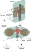
Electrode Positioning to Investigate the Changes of the Thoracic Bioimpedance Caused by Aortic Dissection - A Simulation Study
- PMID: 33584902
- PMCID: PMC7531103
- DOI: 10.2478/joeb-2020-0007
Electrode Positioning to Investigate the Changes of the Thoracic Bioimpedance Caused by Aortic Dissection - A Simulation Study
Abstract
Impedance cardiography (ICG) is a non-invasive method to evaluate several cardiodynamic parameters by measuring the cardiac-synchronous changes in the dynamic transthoracic electrical impedance. ICG allows us to identify and quantify conductivity changes inside the thorax by measuring the impedance on the thorax during a cardiac cycle. Pathologic changes in the aorta, like aortic dissection, will alter the aortic shape as well as the blood flow and consequently, the impedance cardiogram. This fact distorts the evaluated cardiodynamic parameters, but it could lead to the possibility to identify aortic pathology. A 3D numerical simulation model is used to compute the impedance changes on the thorax surface in case of the type B aortic dissection. A sensitivity analysis is applied using this simulation model to investigate the suitability of different electrode configurations considering several patient-specific cases. Results show that the remarkable pathological changes in the aorta caused by aortic dissection alters the impedance cardiogram significantly.
Keywords: Aortic dissection; impedance cardiography; numerical simulation; sensitivity analysis.
© 2020 V. Badeli et al., published by Sciendo.
Conflict of interest statement
Conflict of interest Authors state no conflict of interest.
Figures


References
-
- Khan IA, Nair CK. Clinical, diagnostic and management perspectives of aortic dissection. Elsevier Chest. 2002;122(1):311–28. https://doi.org/10.1378/chest.122.1.311 . - PubMed
-
- Heuser J. Distributed under a CC-BY-SA-3.0 license Wikimedia Commons. 2016.
-
- Patchett N. Distributed under a CC BY-SA 4.0 license. 2015. Wikimedia Commons.
-
- Altamirano-Diaz L, Welisch E, Dempsey AA, Park TS, Grattan M, Norozi K. Non-invasive measurement of cardiac output in children with repaired coarctation of the aorta using electrical cardiometry compared to transthoracic Doppler echocardiography. Physiol Meas. 2018;39(5):055003. 17; https://doi.org/10.1088/1361-6579/aac02b . - PubMed
-
- Reinbacher-Köstinger A, Badeli V, Biro O, Magele C. Numerical simulation of conductivity changes in the human thorax caused by aortic dissection. IEEE Trans. Magnetic. 2019;55(6):5100304. https://doi.org/10.1109/tmag.2019.2895418 .
LinkOut - more resources
Full Text Sources
Other Literature Sources
