

Is Ocular Sonography a Reliable Method for the Assessment of Elevated Intracranial Pressure in Children?
- PMID: 33585057
- PMCID: PMC7870339
- DOI: 10.1055/s-0040-1716385
Is Ocular Sonography a Reliable Method for the Assessment of Elevated Intracranial Pressure in Children?
Abstract
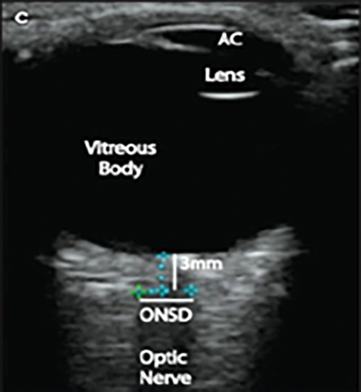
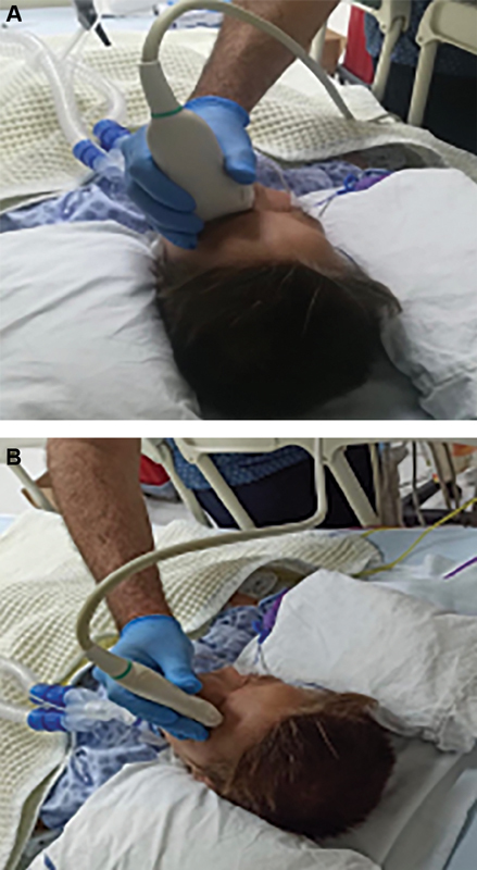
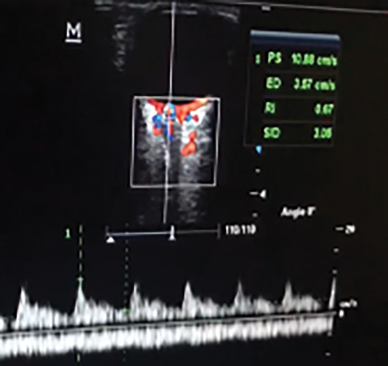
Point-of-care ultrasound has been widely used by clinicians at the bedside in recent years. Various types of point-of-care ultrasound practices are employed, especially in pediatric emergency rooms and intensive care units. Pediatric intensive care specialists perform point-of-care ultrasound virtually as a part of physical examination since it provides just-in-time vital clinical information, which could assist in acute management strategies in critically ill patients. Measurement of optic nerve sheath diameter using point-of-care ultrasound is a noninvasive and radiation-free technique to determine raised intracranial pressure. Ophthalmic artery and central retinal artery Doppler indices can be used as transcranial Doppler to assess raised intracranial pressure. The aim of this review was to provide detailed information on ultrasonographic measurements of optic nerve sheath diameter and central retinal artery Doppler indices as techniques of interest for predicting increased intracranial pressure in pediatric patients in view of the literature.
Keywords: optic nerve sheath diameter; orbital artery Doppler indices; pediatric intensive care unit; point-of-care ultrasound.
Thieme. All rights reserved.
Conflict of interest statement
Conflict of Interest None declared.
Figures
References
-
- O'Brien A J, Brady R M. Point-of-care ultrasound in paediatric emergency medicine. J Paediatr Child Health. 2016;52(02):174–180. - PubMed
-
- Moore C L, Copel J A. Point-of-care ultrasonography. N Engl J Med. 2011;364(08):749–757. - PubMed
-
- Dubourg J, Javouhey E, Geeraerts T, Messerer M, Kassai B. Ultrasonography of optic nerve sheath diameter for detection of raised intracranial pressure: a systematic review and meta-analysis. Intensive Care Med. 2011;37(07):1059–1068. - PubMed
-
- Xirouchaki N, Magkanas E, Vaporidi K. Lung ultrasound in critically ill patients: comparison with bedside chest radiography. Intensive Care Med. 2011;37(09):1488–1493. - PubMed
-
- Longjohn M, Wan J, Joshi V, Pershad J. Point-of-care echocardiography by pediatric emergency physicians. Pediatr Emerg Care. 2011;27(08):693–696. - PubMed
Publication types
LinkOut - more resources
Full Text Sources
