Review
. 2021 Jan 29:10:528624.
doi: 10.3389/fonc.2020.528624. eCollection 2020.

Onco-Esthetics Dilemma: Is There a Role for Electrocosmetic-Medical Devices?

Affiliations
Review

Onco-Esthetics Dilemma: Is There a Role for Electrocosmetic-Medical Devices?

Beniamino Palmieri et al. Front Oncol. .

Abstract

Objective: The primary aim of this review is to verify whether the warning against the use of electromedical instruments in the cosmetic professional or medical cancer patient settings is consistent with evident oncological risks supported by experimental in vitro/in vivo studies or anecdotal clinical reports, or any other reasonable statement.

Methods: MEDLINE, PubMed, Embase, AMED, Ovid, Cochrane Controlled Trials Register, and Google Scholar databases were electronically searched. Data relating to research design, sample population, type of electro-cosmetic devices used, were extracted.

Results: The search strategy identified 50 studies, 30 of which were potentially relevant.

Conclusions: Our research is in favor of moderate periodical use of cosmetic medical devices in patients bearing tumors, in any stage, like in healthy people. Special consideration is dedicated to massage, manipulation, and pressure delivery upon the cytoskeleton of cancer cells that has proven to be sensitive to mechanical stress at least in some specific locally relapsing cancers such as osteosarcoma.

Keywords: cancer; cosmetic; electro-cosmetic device; esthetician; esthetics; medical devices; onco-esthetic; oncology esthetics.


Conflict of interest statement

The authors declare that the research was conducted in the absence of any commercial or financial relationships that could be construed as a potential conflict of interest.

Figures

FIGURE 1
FIGURE 1
(A,B) Gender age-adjusted cancer mortality rates over seven decades since 1930. Adapted from Subra Suresh (Department of Materials Science and Engineering, Division of Biological Engineering, and Harvard-MIT Division of Health Sciences and Technology, Massachusetts Institute of Technology, Cambridge, MA). Copyright 2007 Acta Materialia Inc., Published by Elsevier Ltd., All rights reserved.
FIGURE 2
FIGURE 2
Steps involved in the cancer cell invasion–metastasis cascade. Adapted with modifications from Subra Suresh (Department of Materials Science and Engineering, Division of Biological Engineering, and Harvard-MIT Division of Health Sciences and Technology, Massachusetts Institute of Technology, Cambridge, MA). Copyright 2007 Acta Materialia Inc., Published by Elsevier Ltd., All rights reserved.
FIGURE 3
FIGURE 3
Schematic of chemo biomechanical pathways influencing connections among subcellular structure, cell biomechanics, motility, and disease state. This structure–function–disease paradigm in cancer is a new disease perspective. Copyright 2007 Acta Materialia Inc., Published by Elsevier Ltd., All rights reserved.
FIGURE 4
FIGURE 4
NF-kB signaling pathway. In resting cells, the dimeric transcription factor NF-kB, composed of p50 and p65, is sequestered in the cytosol, bound to the inhibitor I-kB. Stimulation by TNF-α or IL-1 induces activation of TAK1 kinase (step 1), leading to activation of the trimeric I-kB kinase (step 2a). Ionizing radiation and other stresses can directly activate I-kB kinase by an unknown mechanism (step 2b). Following phosphorylation of I-kB by I-kB kinase and binding of E3 ubiquitin ligase (step 3), polyubiquitination of I-kB (step 4) targets is for degradation by proteasomes (step 5). The removal of I-kB unmasks the nuclear-localization signals (NLS) in both subunits of NF-kB, allowing their translocation to the nucleus. Copyright 2011 Receptors and signal transduction Published by Technology. All rights reserved.
FIGURE 5
FIGURE 5
Schematic illustration of the subcellular structure of a typical eukaryotic cell. Adapted from Subra Suresh (Department of Materials Science and Engineering, Division of Biological Engineering, and Harvard-MIT Division of Health Sciences and Technology, Massachusetts Institute of Technology, Cambridge, MA). Copyright 2007 Acta Materialia Inc., Published by Elsevier Ltd., All rights reserved.
FIGURE 6
FIGURE 6
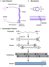
Description of the components of the cytoskeleton: structure of (A) actin microfilament (polymer that plays a key role in muscle contraction, cell movement and shape), (B) microtubule (long tubulin filament that plays a role in cell structure, organization, mitosis, and movement) and (C) intermediate filament (ropelike cytoskeletal filament that plays several different structural roles but is not involved in cell movement). Reproduced with permission from Subra Suresh (Department of Materials Science and Engineering, Division of Biological Engineering, and Harvard-MIT Division of Health Sciences and Technology, Massachusetts Institute of Technology, Cambridge, MA). Copyright 2007 Acta Materialia Inc., Published by Elsevier Ltd., All rights reserved.
FIGURE 7
FIGURE 7
Schematic illustrations of the biomechanical assays on tumor cells (A–C). Biophysical assays commonly used to probe the deformation of single cells are illustrated in (D–G). Techniques used to infer cytoadherence, deformation, and mobility characteristics of populations of cells are schematically sketched in (H,I). Image courtesy of, and with permission from, Subra Suresh (Department of Materials Science and Engineering, Division of Biological Engineering, and Harvard-MIT Division of Health Sciences and Technology, Massachusetts Institute of Technology, Cambridge, MA). Copyright 2007 Acta Materialia Inc., Published by Elsevier Ltd., All rights reserved.
FIGURE 8
FIGURE 8
Working model of the mechanical response pathway by which pressure and shear stimulate tumor cell adhesion. FAK is activated in response to mechanical stimuli through a dual signaling pathway requiring an intact cytoskeleton, α-actinin, and paxillin as well as cytoskeleton-independent activation of Src. FAK and Src colocalize to β1-integrin heterodimers in an Akt- and α-actinin-dependent manner, respectively. FAK–Src complex formation and signaling stimulates PI3K-dependent activation of ILK, resulting in β1-integrin phosphorylation and conformational activation, increasing substrate binding affinity. Image courtesy of, and with permission from, Ref. David H. Craig (Department of Surgery; Michigan State University; Lansing, Michigan United States). Copyright 2009 Cell Cycle Published by HHS Public Access. All rights reserved.
FIGURE 9
FIGURE 9
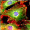
A mouse NIH3T3 fibroblast cell was fixed and stained for DNA (blue) and the major cytoskeletal filaments actin (red) and alpha-tubulin (green). The cell was imaged by fluorescence microscopy on an optical IX70 microscope with a deep-cooled CCD camera. Image courtesy of, and with permission from, Andrew E. Pelling (London Centre for Nanotechnology and Department of Medicine, University College London). Copyright 2007 Acta Materialia Inc., Published by Elsevier Ltd., All rights reserved.
FIGURE 10
FIGURE 10
Talin structure and binding partners. (A) Relationship between talin and its signaling/structural partners. (B) Structure of the talin integrin-binding FERM domain head and mechanosensitive rod domain, with binding site locations. PIP2, phosphatidylinositol 4,5-bisphosphate. Adapted from Ref. (18). Copyright 1987 John Wiley & Sons – Journals. Published by Federation of American Societies for Experimental Biology. All rights reserved.
FIGURE 11
FIGURE 11
The role of talin in malignant phenotypes. Adapted from Ref. (18). Copyright 1987 John Wiley & Sons—Journals. Published by Federation of American Societies for Experimental Biology. All rights reserved.

Comment in

References

    1. Hanahan D, Weinberg RA. Hallmarks of Cancer: the next generation. Cell. (2011) 144:646–74. 10.1016/j.cell.2011.02.013 - DOI - PubMed
    1. Shen R, Ye Y, Chen L, Yan Q, Barsky SH, Gao JX. Precancerous stem cells can serve as tumor vasculogenic progenitors. PLoS One. (2008) 3:e1652. 10.1371/journal.pone.0001652 - DOI - PMC - PubMed
    1. Chu WM. Tumor necrosis factor. Cancer Lett. (2013) 328:222–5. - PMC - PubMed
    1. Lou J, Lucas R, Grau GE. Pathogenesis of cerebral malaria: recent experimental data and possible applications for humans. Clin Microbiol Rev. (2001) 14:810–20. 10.1128/CMR.14.4.810-820.2001 - DOI - PMC - PubMed
    1. Institute of Medicine (US) Committee on Assessing Interactions Among Social, Behavioral, and Genetic Factors in Health, Hernandez LM, Blazer DG. Genes, Behavior, and the Social Environment: Moving Beyond the Nature/Nurture Debate. Washington, DC: National Academies Press. (2006). 10.1128/cmr.14.4.810-820.2001 - DOI - PubMed

LinkOut - more resources