

Is Senescence-Associated β-Galactosidase a Reliable in vivo Marker of Cellular Senescence During Embryonic Development?
- PMID: 33585480
- PMCID: PMC7876289
- DOI: 10.3389/fcell.2021.623175
Is Senescence-Associated β-Galactosidase a Reliable in vivo Marker of Cellular Senescence During Embryonic Development?
Abstract
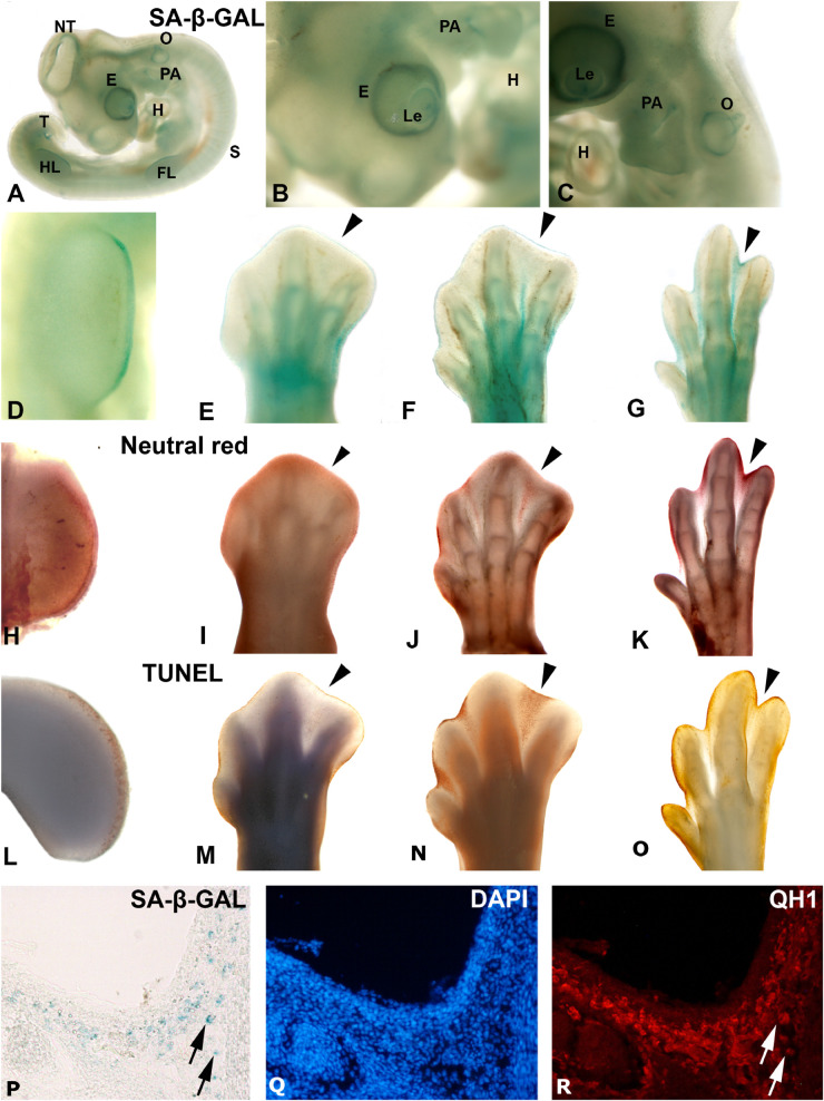
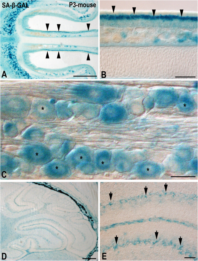
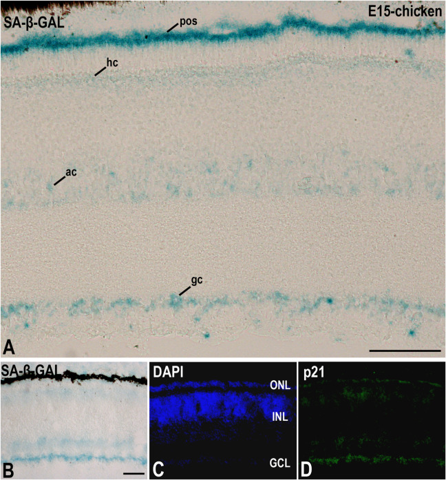
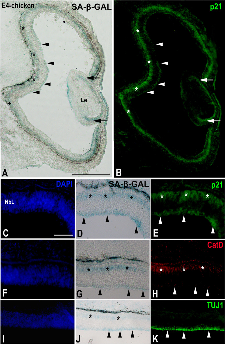
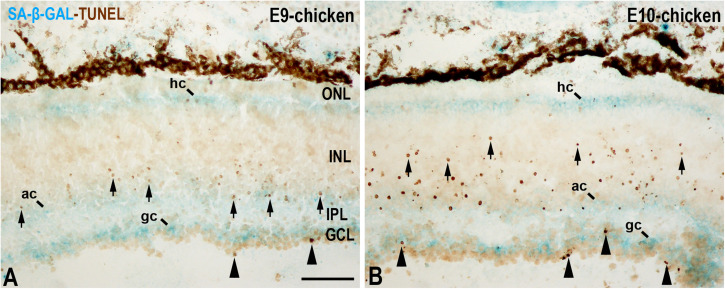
During vertebrate embryonic development, cellular senescence occurs at multiple locations. To date, it has been accepted that when there has been induction of senescence in an embryonic tissue, β-galactosidase activity is detectable at a pH as high as 6.0, and this has been extensively used as a marker of cellular senescence in vivo in both whole-mount and cryosections. Such senescence-associated β-galactosidase (SA-β-GAL) labeling appears enhanced in degenerating regions of the vertebrate embryo that are also affected by programmed cell death. In this sense, there is a strong SA-β-GAL signal which overlaps with the pattern of cell death in the interdigital tissue of the developing limbs, and indeed, many of the labeled cells detected go on to subsequently undergo apoptosis. However, it has been reported that β-GAL activity at pH 6.0 is also enhanced in healthy neurons, and some retinal neurons are strongly labeled with this histochemical technique when they begin to differentiate during early embryonic development. These labeled early post-mitotic neurons also express other senescence markers such as p21. Therefore, the reliability of this histochemical technique in studying senescence in cells such as neurons that undergo prolonged and irreversible cell-cycle arrest is questionable because it is also expressed in healthy post-mitotic cells. The identification of new biomarkers of cellular senescence would, in combination with established markers, increase the specificity and efficiency of detecting cellular senescence in embryonic and healthy mature tissues.
Keywords: cell death; cell senescence; development; histochemistry; limb; retina.
Copyright © 2021 de Mera-Rodríguez, Álvarez-Hernán, Gañán, Martín-Partido, Rodríguez-León and Francisco-Morcillo.
Conflict of interest statement
The authors declare that the research was conducted in the absence of any commercial or financial relationships that could be construed as a potential conflict of interest.
Figures
References
-
- Ahuja S., Ahuja-Jensen P., Johnson L. E., Caffé A. R., Abrahamson M., Ekström P. A. R., et al. (2008). Rd1 mouse retina shows an imbalance in the activity of cysteine protease cathepsins and their endogenous inhibitor cystatin. Investig. Ophthalmol. Vis. Sci. 49 1089–1096. 10.1167/iovs.07-0549 - DOI - PubMed
-
- Álvarez-Hernán G., Andrade J. P., Escarabajal-Blázquez L., Blasco M., Solana-Fajardo J., Martín-Partido G., et al. (2019). Retinal differentiation in syngnathids: comparison in the developmental rate and acquisition of retinal structures in altricial and precocial fish species. Zoomorphology 138 371–385. 10.1007/s00435-019-00447-3 - DOI
Publication types
LinkOut - more resources
Full Text Sources
Other Literature Sources