

Optical Coherence Tomography Angiography Findings After Intravitreal Ranibizumab in Patients With Coats Disease
- PMID: 33585512
- PMCID: PMC7873908
- DOI: 10.3389/fmed.2020.615015
Optical Coherence Tomography Angiography Findings After Intravitreal Ranibizumab in Patients With Coats Disease
Abstract
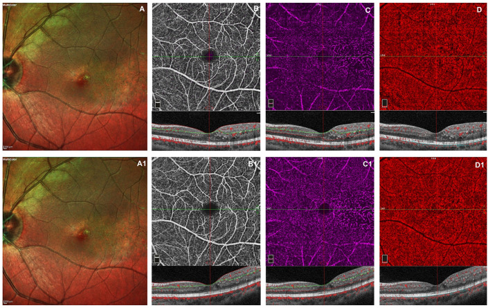
The aim of this retrospective study was to describe the vascular features in eyes with Coats disease, using optical coherence tomography angiography (OCTA), at baseline and after 3 monthly intravitreal injections of ranibizumab. Fifteen eyes of 15 consecutive patients affected by Coats' disease were recruited in this study. All patients underwent the best-corrected visual acuity (BCVA) evaluation, fundus examination, fluorescein angiography (FA), indocyanine green angiography (ICGA), multicolor imaging, structural Spectral Domain (SD)-OCT and OCTA at baseline and 1 month after the third monthly ranibizumab injection (loading phase). Fifteen patients completed the study, of whom nine were males and six females. Mean age was 20.4 ± 2 years. BCVA was 0.46 ± 0.11 logMar and 0.47 ± 0.12 logMar at baseline and after treatment, respectively (p = 0.164). SD-OCT revealed no significant decrease in central macular thickness (486.33 μm ± 93.37 at baseline vs. 483.4 μm ± 80.97 after treatment; p = 0.915). The subretinal exudates persisted in macular region after intravitreal injections. OCTA showed a general vascular rarefaction in superficial capillary plexus (SCP), deep capillary plexus (DCP), and choriocapillary (CC) that did not change after loading phase. This study showed no functional and vascular improvement following 3 monthly ranibizumab injections. OCTA, non-invasive technique, could be useful during follow up of these patients and provide a better understand of pathogenesis of this disorder.
Keywords: Coats disease; OCTA; SD-OCT; anti-VEGF injections; retinal vascular features.
Copyright © 2021 Cennamo, Montorio, Comune, Laezza, Fallico, Lionetti and Reibaldi.
Conflict of interest statement
The authors declare that the research was conducted in the absence of any commercial or financial relationships that could be construed as a potential conflict of interest.
Figures
References
-
- Coats G. Forms of retinal diseases with massive exudation. R Lond Ophthalmol Hosp Rep. (1908) 17:440e525
LinkOut - more resources
Full Text Sources
Other Literature Sources
Miscellaneous