. 2021 Jan 27:7:617038.
doi: 10.3389/fcvm.2020.617038. eCollection 2020.

Cardiomyocyte Na+/H+ Exchanger-1 Activity Is Reduced in Hypoxia

Affiliations

Cardiomyocyte Na+/H+ Exchanger-1 Activity Is Reduced in Hypoxia

Hilmi Burak Kandilci et al. Front Cardiovasc Med. .

Abstract

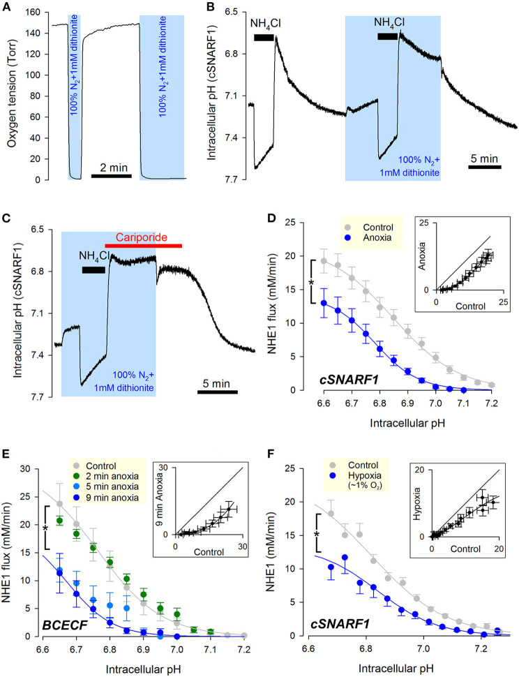
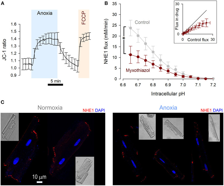
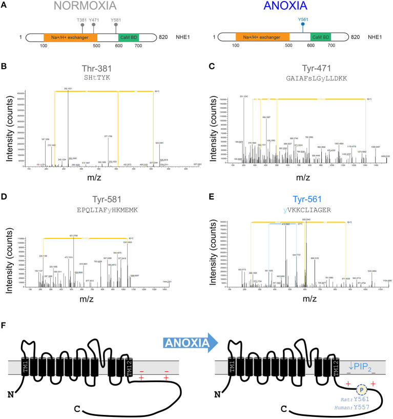
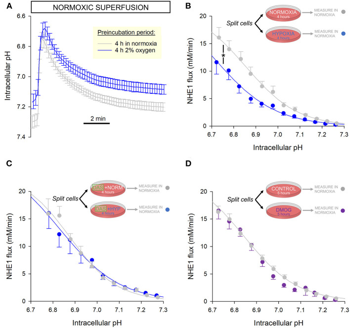
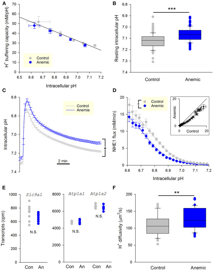
Fully-activated Na+/H+ exchanger-1 (NHE1) generates the cardiomyocyte's largest trans-membrane extrusion of H+ ions for an equimolar influx of Na+ ions. This has the desirable effect of clearing excess intracellular acidity, but comes at a large energetic premium because the exchanged Na+ ions must ultimately be extruded by the sodium pump, a process that consumes the majority of the heart's non-contractile ATP. We hypothesize that the state of NHE1 activation depends on metabolic resources, which become limiting in periods of myocardial hypoxia. To test this functionally, NHE1 activity was measured in response to in vitro and in vivo hypoxic treatments. NHE1 flux was interrogated as a function of intracellular pH by fluorescence imaging of rodent ventricular myocytes loaded with pH-sensitive dyes BCECF or cSNARF1. Anoxic superfusates promptly inhibited NHE1, tracking the time-course of mitochondrial depolarization. Mass spectrometry of NHE1 immuno-precipitated from Langendorff-perfused anoxic hearts identified Tyr-581 dephosphorylation and Tyr-561 phosphorylation. The latter residue is part of the domain that interacts with phosphatidylinositol 4,5-bisphosphate (PIP2), a membrane lipid that becomes depleted under metabolic inhibition. Tyr-561 phosphorylation is expected to electrostatically weaken this activatory interaction. To test if a period of hypoxia produces a persistent inhibition of NHE1, measurements under normoxia were performed on myocytes that had been incubated in 2% O2 for 4 h. NHE1 activity remained inhibited, but the effect was ablated in the presence of Dasatinib, an inhibitor of Abl/Src-family tyrosine kinases. Chronic tissue hypoxia in vivo, attained in a mouse model of anemic hypoxia, also resulted in persistently slower NHE1. In summary, we show that NHE1 responds to oxygen, a physiologically-relevant metabolic regulator, ostensibly to divert ATP for contraction. We describe a novel mechanism of NHE1 inhibition that may be relevant in cardiac disorders featuring altered oxygen metabolism, such as myocardial ischemia and reperfusion injury.

Keywords: ATP; NHE1; Y557; anemia; metabolism; oxygen; pH regulation; ventricle.


Conflict of interest statement

YC has received personal support from Vifor Pharma for work on iron biology not related to the current study. The remaining authors declare that the research was conducted in the absence of any commercial or financial relationships that could be construed as a potential conflict of interest.

Figures

Figure 1
Figure 1
Cardiac NHE1 activity is rapidly inhibited by acute anoxia. (A) Measurement of oxygen tension in the superfusion chamber, showing anoxic conditions are attainable by solutions bubbled with N2 and supplemented with the oxygen-scavenger dithionite (1 mM). (B) Exemplar trace of paired ammonium prepulses, first performed in normoxia and then in anoxia, in a rat ventricular myocyte loaded with cSNARF1. Recovery of pHi from the acid load is greatly attenuated under anoxic conditions, but becomes re-activated upon re-oxygenation. (C) Exemplar time course showing lack of background acid-loading at low pHi under anoxic conditions, revealed in the presence of cariporide (30 μM) to block NHE1. (D) NHE1-generated H+-flux measured by cSNARF1 or (E) BCECF in normoxia (cSNARF1: n = 22 cells from 5 rats; BCECF: n = 9 cells from 4 rats) or 9 min anoxia (cSNARF: n = 9 cells from 2 rats; BCECF: n = 7 cells from 2 rats). In the case of BCECF experiments, the effect of anoxia was also tested after 2 min exposure (n = 5 cells from 3 rats), 5 min exposure to anoxia (n = 5 cells from 3 rats). Two-way ANOVA: effect of 9 min anoxia vs. control was significant (P < 0.0001 for cSNARF1 and P < 0.01 for BCECF datasets); effect of 5 min anoxia vs. control was significant (P < 0.0001); effect of 2 min anoxia was not significant (P = 0.93). Interaction between pH and treatment was not significant in any of the cases; the effect of pHi was significant (P < 0.0001 for all datasets). Insets plot flux in anoxia (9 min) vs. normoxia for matching pHi; the parallel shift is indicative of a change in affinity. (F) Effect of 15 min 3 Torr hypoxia (~1% O2) attained by including 0.1 mM dithionite in N2-bubbled solutions. Data recorded with cSNARF1. NHE1 activity was inhibited by hypoxia (n = 26 cells, 3 rats) relative to controls (n = 55, 3 rats). Two-way ANOVA: significant effect of hypoxia (P < 0.001); of pHi (P < 0.0001); and interaction (P < 0.0001). Inset plots flux in anoxia vs. normoxia for matching pHi. The observed parallel shift is indicative of a change in affinity.
Figure 2
Figure 2
NHE1 activity is reduced by metabolic inhibition. (A) JC-1 fluorescence ratio measured in rat ventricular myocytes (n = 12 cells from 4 rats), showing mitochondrial depolarization in response to anoxic conditions. The response was comparable to the effect of FCCP (5 μM), indicating a complete depolarization by anoxia. (B) NHE1-generated flux measured in ventricular myocytes in the presence myxothiazol (1 μM, n = 7 cells from 3 rats), compared to controls. Two-way ANOVA: significant effect of drug (P < 0.0001); of pHi (P < 0.0001); and interaction (P = 0.0461). Inset plots flux in the presence and absence of inhibitor for matching pHi. (C) Immunofluorescence of rat ventricular myocytes for NHE1 under control conditions and after treatment with anoxic conditions for 15 min. NHE1 distribution appears unaltered. Scale bar applies to both images.
Figure 3
Figure 3
Anoxia changes the phosphorylation pattern at the NHE1 C-terminus. (A) Phosphorylation sites identified by tandem mass spectrometry-based proteomics analysis on NHE1 pulled-down from rat hearts Langendorff-perfused with either anoxic (n = 4) or normoxic (n = 4; control) buffers for 15 min prior to sample collection. Threonine residue at position 381 (T381) and tyrosine residues at positions 471 (Y471) and 581 (Y581) were identified in normoxia conditions, while tyrosine residue at position 561 (Y561) was identified in hypoxia conditions only. The Na+/H+ exchanger region and calmodulin binding domain (Cam BD) are highlighted in orange and green, respectively, on NHE1 protein. (B–D) Tandem mass spectra containing phosphorylated T381, Y471, Y581, and Y561 residues matching the corresponding peptide ions in (B) SHTTIK (z = +2, m/z = 383.68954), (C) GAIAFSLGYLLDKK (z = +2, m/z = 849.36743), (D) EPQLIAFYHKMEMK (z = +3, m/z = 615.64807) and (E) YVKKCLIAGER (z = +3, m/z = 472.89618) are shown. (F) Schematic of NHE1 protein structure, showing positively charged lipid interacting domain of the C-terminus. Anoxia leads to phosphorylation which weakens the otherwise activatory interaction with negatively charged PIP2 in the inner leaf of the sarcolemma. Additionally, metabolic inhibition will deplete PIP2 levels.
Figure 4
Figure 4
NHE1 activity is persistently inhibited following 4 h incubation in hypoxia. (A) Freshly isolated rat myocytes were split and incubated either in an atmosphere of 2% O2 or normal air. Low O2 was maintained by under an Ibidi Chamber. After incubation, NHE1 activity was measured in myocytes under normoxic superfusion, within 30 min of re-oxygenation (n = 30 control cells and 28 hypoxia-pretreated cells from 4 rats). (B) NHE1 activity remained lower in cells that had been exposed to hypoxia, indicating that the inhibitory effect of low O2 persists after re-oxygenation. Two-way ANOVA: significant effect of treatment (hypoxic pre-incubation) (P < 0.0001) and interaction between treatment and pH (P < 0.0001). (C) Experiments were repeated in the presence of 100 nM Dasatinib, a Abl/Src tyrosine kinase inhibitor. In the presence of inhibitor, hypoxia had no effect on NHE1, consistent with the role of Y561 phosphorylation (n = 15 controls cells, 16 hypoxia pre-treated cells; from 3 rats); two-way ANOVA: no significant effect of treatment (hypoxic pre-incubation) in presence of Dasatinib (DAS) (P = 0.96); no significant interaction with pH (P = 0.98). (D) 5 h treatment with 1 mM DMOG had no effect on NHE1 in rat myocytes, compared to controls (n = 34 controls cells, 27 DMOG-treated cells from 4 animals); two-way ANOVA: no significant effect of treatment (DMOG) (P = 0.78).
Figure 5
Figure 5
Chronic myocardial hypoxia in anemic animals persistently reduces NHE1 activity. (A) Intrinsic buffering capacity in myocytes from control (n = 20 cells, 3 mice) and anemic mice (n = 23 cells, 3 mice). No significant difference (two-way ANOVA). (B) Resting pHi under Hepes-buffering regime (n = 124 and 121 cells from 3 anemic and 3 control mice, respectively); t-test, P < 0.01. (C) Time course of pHi recovery following an ammonium prepulse. Average of 46 myocytes from three control hearts and 36 myocytes from three anemic hearts. Error bars not shown for clarity. (D) NHE1 generated flux in myocytes from control (n = 46 cells from 3 mice) and anemic (n = 36 cells from 3 mice) animals, showing significant decrease in the latter; two-way ANOVA: significant (P < 0.01) effect of anemic intervention. (E) Levels of transcripts for the gene coding NHE1, Slc9a1, is not different between anemic and control hearts. Whole heart tissue mRNA from 9 mice per group. Used as a control, Na+/K+ pump genes Atp1a1 and Atp1a2 are unchanged. (F) Apparent H+ diffusion coefficient is significantly increased in myocytes from anemic mice (n = 12 from 3 mice) relative to control myocytes (n = 12 from 3 mice); t-test, P < 0.01.

Similar articles

Cited by

References

    1. Pedersen SF, Counillon L. The SLC9A-C mammalian Na(+)/H(+) exchanger family: molecules, mechanisms, and physiology. Physiol Rev. (2019) 99:2015–113. 10.1152/physrev.00028.2018 - DOI - PubMed
    1. Orlowski J, Grinstein S. Na+/H+ exchangers of mammalian cells. J Biol Chem. (1997) 272:22373–6. 10.1074/jbc.272.36.22373 - DOI - PubMed
    1. Vaughan-Jones RD, Spitzer KW, Swietach P. Intracellular pH regulation in heart. J Mol Cell Cardiol. (2009) 46:318–31. 10.1016/j.yjmcc.2008.10.024 - DOI - PubMed
    1. Garciarena CD, Youm JB, Swietach P, Vaughan-Jones RD. H(+)-activated Na(+) influx in the ventricular myocyte couples Ca(2)(+)-signalling to intracellular pH. J Mol Cell Cardiol. (2013) 61:51–9. 10.1016/j.yjmcc.2013.04.008 - DOI - PubMed
    1. Suga H. Ventricular energetics. Physiol Rev. (1990) 70:247–77. 10.1152/physrev.1990.70.2.247 - DOI - PubMed

LinkOut - more resources